. 2011 Oct;39(18):7931-45.
doi: 10.1093/nar/gkr523. Epub 2011 Jul 3.

ASF1A and ATM regulate H3K56-mediated cell-cycle checkpoint recovery in response to UV irradiation

Affiliations

ASF1A and ATM regulate H3K56-mediated cell-cycle checkpoint recovery in response to UV irradiation

Aruna Battu et al. Nucleic Acids Res. 2011 Oct.

Abstract

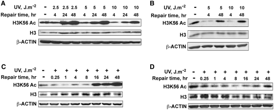
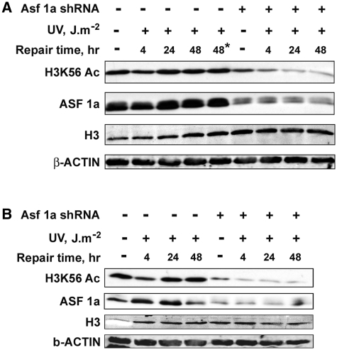
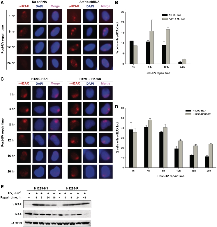
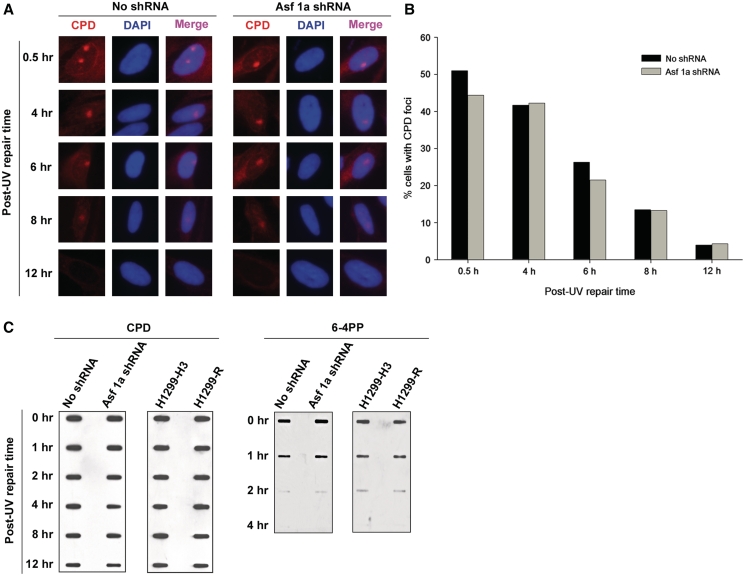
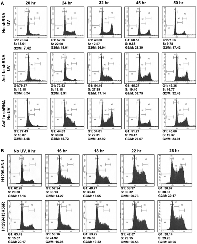
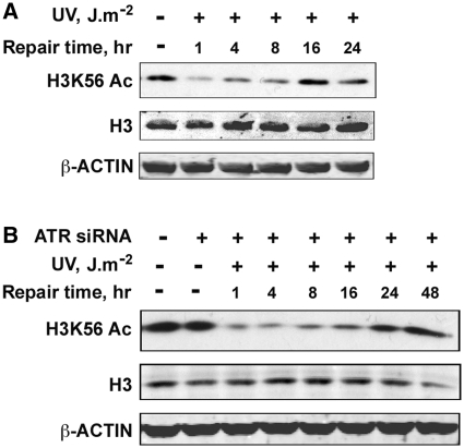
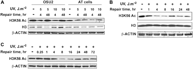
Successful DNA repair within chromatin requires coordinated interplay of histone modifications, chaperones and remodelers for allowing access of repair and checkpoint machineries to damaged sites. Upon completion of repair, ordered restoration of chromatin structure and key epigenetic marks herald the cell's normal function. Here, we demonstrate such a restoration role of H3K56 acetylation (H3K56Ac) mark in response to ultraviolet (UV) irradiation of human cells. A fast initial deacetylation of H3K56 is followed by full renewal of an acetylated state at ~24-48 h post-irradiation. Histone chaperone, anti-silencing function-1 A (ASF1A), is crucial for post-repair H3K56Ac restoration, which in turn, is needed for the dephosphorylation of γ-H2AX and cellular recovery from checkpoint arrest. On the other hand, completion of DNA damage repair is not dependent on ASF1A or H3K56Ac. H3K56Ac restoration is regulated by ataxia telangiectasia mutated (ATM) checkpoint kinase. These cross-talking molecular cellular events reveal the important pathway components influencing the regulatory function of H3K56Ac in the recovery from UV-induced checkpoint arrest.


Figures

Figure 1.
Figure 1.
H3K56 is deacetylated in response to UV irradiation and acetylation is promptly restored after several hours of post-repair. (A) HeLa cells were exposed to increasing doses (2.5, 5 and 10 J m−2) of UV radiation, whole-cell lysates prepared immediately or at the indicated times, and the H3K56Ac levels determined by western blotting. (B) OSU-2 NHF cells were exposed to 5 and 10 J m−2 UV and cell lysates were prepared at the indicated post-repair times. Equal amounts of cell lysates were resolved on SDS–PAGE and probed for H3K56Ac levels by western blotting. (C) HeLa cells were exposed to 10 J m−2 UV irradiation, cell lysates prepared at various post-repair times and H3K56Ac levels determined by western blotting. (D) OSU-2 cells were exposed to 10 J m−2 UV irradiation and processed as described above for (C). Total H3 and β-actin levels were determined to ascertain equal protein loading.
Figure 2.
Figure 2.
Restoration of H3K56 acetylation after UV damage repair is regulated by ASF1A histone chaperone. (A) ASF1A is required for H3K56Ac restoration in asynchronous NHF cells after UV damage repair. OSU-2 cells were transfected with ASF1A shRNA and after 24 h irradiated at 10 J m−2 UV. Cultures were again transfected with ASF1A shRNA to maintain the knockdown up to 48 h post-irradiation. Whole cell lysates were prepared at the indicated times and H3K56Ac levels determined by western blotting. Asterik indicates cells treated with Fugene transfection reagent alone to exclude any possible toxicity effects on H3K56Ac levels. (B) Regulation of H3K56Ac restoration by ASF1A in response to UV irradiation is independent of the effects of ASF1A during replication. OSU-2 cells were arrested in the G1 phase of the cell-cycle by serum starvation for 48 h, transfected with ASF1A shRNA and exposed to 10 J m−2 UV irradiation after 24 h of transfection. Cells were transfected again with ASF1A shRNA after UV irradiation to maintain the knockdown for a further 48 h. Cell lysates were prepared immediately or at the indicated post-UV repair times. H3K56Ac levels were determined by western blotting using anti-H3K56Ac antibody.
Figure 3.
Figure 3.
H3K56 acetylation is required for the dephosphorylation of γ-H2AX. (A) Depletion of ASF1A results in the retention of γ-H2AX for a longer time at sites of UV damage. OSU-2 cells were transfected with ASF1A shRNA and after 24 h irradiated with 10 J m−2 UV through micro-pore filters placed on the cell monolayers. Cells were recovered at indicated times and fixed with paraformaldehyde. Immunofluorescence was performed using anti-γ-H2AX antibody. Images shown are from a representative of multiple experiments. (B) Quantitation of γ-H2AX foci in ASF1A-depleted cells. Triplicate experiments were performed as described above in (A) and the number of cells containing γ-H2AX foci in shRNA-treated and -untreated cells were quantitated as follows. An average of 150 merged nuclear foci from at least five different microscopic fields were used for the quantitation of γ-H2AX foci for each experiment. The graph depicts the average number of cells containing γ-H2AX foci from the three independent experiments. The error bars show the mean standard deviation. (C) H3K56R mutation results in the retention of γ-H2AX foci at sites of UV damage. H1299-H3.1 and H1299-H3K56R cells were exposed to 10 J m−2 of UV radiation through micro-pore filters. Cells were fixed at the indicated intervals and immunofluorescence was performed to detect γ-H2AX foci. Images shown are from a representative experiment. (D) Quantitation of γ-H2AX foci in H1299 cells. Quantitation of γ-H2AX foci in H1299-H3.1 and H1299-H3K56R cells was essentially performed as described in (B) from two independent experiments. (E) H3K56 acetylation-deficient cells are defective in γ-H2AX dephosphorylation. H1299-H3.1 and H1299-H3K56R cells were irradiated at 10 J m−2 UV and whole cell lysates prepared at 4, 8, 24 and 48 h. Equal quantities of cell lysates were resolved on SDS–PAGE and the levels of γ-H2AX determined by western blotting. Total H2AX and β-ACTIN were used as loading controls.
Figure 4.
Figure 4.
Acetylation of H3K56 is not required for NER. (A) Depletion of ASF1A does not affect CPD repair efficiency. OSU-2 cells were transfected with ASF1A shRNA and irradiated with 10 J m−2 UV after 24 h through micropore filters placed on the cell monolayers. Cells were fixed with paraformaldehyde at the indicated times and immunofluorescence was performed using anti-CPD antibody. Images shown are from a representative of duplicate experiments. (B) Quantitation of CPD foci in ASF1A-depleted cells. Duplicate experiments were performed as described above in (A) and the number of cells containing CPD foci in shRNA-treated and -untreated cells were quantitated as follows. An average of 150 merged nuclear foci from at least five different microscopic fields were used for the quantitation of CPD foci for each experiment. The graph depicts the average number of cells containing CPD foci from two independent experiments. (C) ASF1A-depleted and H1299-H3K56R cells are proficient in NER. ASF1A shRNA-treated cells and H1299-H3K56R cells were irradiated at 10 J m−2 UV and DNA isolated at the indicated post-irradiation times. Equal amounts of DNA was blotted on nitrocellulose membranes and probed for the presence of CPD and 6-4PP photoproducts with specific antibodies.
Figure 5.
Figure 5.
Restoration of H3K56 acetylation is necessary for recovery from UV-induced cell-cycle checkpoint arrest. (A) ASF1A-deficient cells exhibit cell-cycle checkpoint recovery defect. OSU-2 cells were arrested in G1 phase of the cell-cycle by serum starvation for 48 h. The arrested cells were transfected with ASF1A shRNA and after 24 h irradiated with 5 J m−2 UV and released into the cell-cycle by addition of media containing serum. Transfection with ASF1A shRNA was performed again to maintain the knockdown up to 50 h. Cells were harvested at the indicated times, fixed with 70% ethanol and the DNA content determined by FACS. (B) H3K56R mutant cells are defective in recovery from UV-induced cell-cycle checkpoint arrest. H1299-H3.1 and H1299-H3K56R cells were synchronized by serum starvation for 24 h. Cells were then irradiated with 10 J m−2 UV and released into the cell-cycle by addition of media containing serum. Cells were harvested at the indicated time points, fixed and DNA content determined by FACS.
Figure 6.
Figure 6.
H3K56 deacetylation and acetylation is not regulated by ATR checkpoint kinase. (A) Seckel cells are proficient in the deacetylation and restoration of H3K56 acetylation in response to UV irradiation. Seckel cells were exposed to 10 J m−2 UV radiation and were harvested at the indicated times. Whole cell lysates prepared from the cells were resolved on SDS–PAGE and H3K56 acetylation levels determined by western blotting. (B) ATR deficiency does not affect either the deacetylation or restoration of H3K56 acetylation in response to UV irradiation. HeLa cells were transfected with 100 nM ATR siRNA using Lipofectamine tansfection reagent. At 48 h after transfection, cells were irradiated with 10 J m−2 UV and harvested at the indicated times. Whole-cell lysates were prepared, resolved on SDS–PAGE and H3K56Ac levels determined by western blotting with anti-H3K56Ac antibodies.
Figure 7.
Figure 7.
Restoration of H3K56 acetylation is regulated by ATM checkpoint kinase. (A) AT cells are deficient in the restoration of H3K56 acetylation after UV repair. OSU-2 and AT cells were irradiated with 5 and 10 J m−2 UV, allowed to repair for 4 and 48 h and harvested to prepare whole-cell lysates. H3K56 acetylation levels were determined by western blotting. (B) AT cells are deficient in the restoration of H3K56 acetylation, but not the deacetylation in response to UV irradiation. AT cells were irradiated with 10 J m−2 UV and cells harvested at various intervals of post-UV repair. Whole-cell lysates were prepared and H3K56Ac levels analyzed by western blotting. (C) Depletion of ATM results in defective restoration of H3K56 acetylation. HeLa cells stably transfected with ATM shRNA were irradiated with 10 J m−2 UV, allowed to repair and harvested at various times. Whole-cell lysates were prepared and H3K56Ac levels determined by western blotting.
Figure 8.
Figure 8.
A model depicting the role of H3K56 deacetylation/acetylation in DNA damage response.

References

    1. Harper JW, Elledge SJ. The DNA damage response: ten years after. Mol. Cell. 2007;28:739–745. - PubMed
    1. Green CM, Almouzni G. When repair meets chromatin. First in series on chromatin dynamics. EMBO Rep. 2002;3:28–33. - PMC - PubMed
    1. De Boer J, Hoeijmakers JH. Nucleotide excision repair and human syndromes. car. 2000;21:453–460. - PubMed
    1. Reardon JT, Sancar A. Nucleotide excision repair. Prog. Nucleic Acid Res. Mol. Biol. 2005;79:183–235. - PubMed
    1. Mitchell DL, Haipek CA, Clarkson JM. Photoproducts are removed from the DNA of UV-irradiated mammalian cells more efficiently than cyclobutane pyrimidine dimers. Mutat. Res. 1985;143:109–112. - PubMed

Publication types

MeSH terms