. 2011 Aug 30;47(2):167-77.
doi: 10.1530/JME-11-0066. Print 2011 Oct.

Prolonged exposure to GH impairs insulin signaling in the heart

Affiliations

Prolonged exposure to GH impairs insulin signaling in the heart

J G Miquet et al. J Mol Endocrinol. .

Abstract

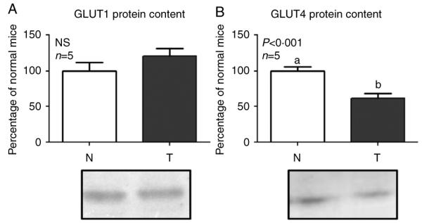
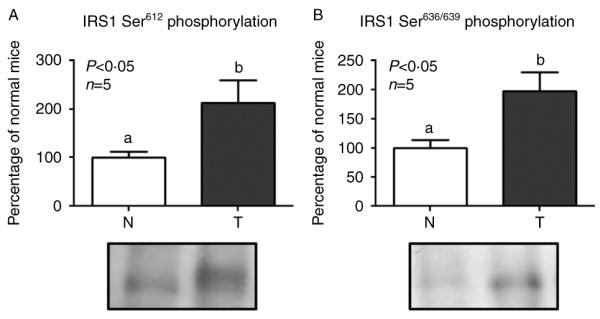
Acromegaly is associated with cardiac hypertrophy, which is believed to be a direct consequence of chronically elevated GH and IGF1. Given that insulin is important for cardiac growth and function, and considering that GH excess induces hyperinsulinemia, insulin resistance, and cardiac alterations, it is of interest to study insulin sensitivity in this tissue under chronic conditions of elevated GH. Transgenic mice overexpressing GH present cardiomegaly and perivascular and interstitial fibrosis in the heart. Mice received an insulin injection, the heart was removed after 2 min, and immunoblotting assays of tissue extracts were performed to evaluate the activation and abundance of insulin-signaling mediators. Insulin-induced tyrosine phosphorylation of the insulin receptor (IR) was conserved in transgenic mice, but the phosphorylation of IR substrate 1 (IRS1), its association with the regulatory subunit of the phosphatidylinositol 3-kinase (PI3K), and the phosphorylation of AKT were decreased. In addition, total content of the glucose transporter GLUT4 was reduced in transgenic mice. Insulin failed to induce the phosphorylation of the mammalian target of rapamycin (mTOR). However, transgenic mice displayed increased basal activation of the IR/IRS1/PI3K/AKT/mTOR and p38 signaling pathways along with higher serine phosphorylation of IRS1, which is recognized as an inhibitory modification. We conclude that GH-overexpressing mice exhibit basal activation of insulin signaling but decreased sensitivity to acute insulin stimulation at several signaling steps downstream of the IR in the heart. These alterations may be associated with the cardiac pathology observed in these animals.


Figures

Figure 1
Figure 1
Myocardial fibrosis evaluation by Masson’s trichrome staining. Representative histological sections showing in blue staining the cardiac extracellular matrix content in normal (N) (A and B) and GH transgenic (T) (C and D) mice (400×). The percentage of blue-stained area corresponding to fibrosis in relation to the total heart area is shown in panel E. Data are the mean±s.e.m., three different individuals of each group were analyzed in parallel. Groups denoted by different letters are significantly different, P<0·05. Full colour version of this is figure available via http://dx.doi.org/10.1530/JME-11-0066.
Figure 2
Figure 2
Insulin receptor (IR) and insulin receptor substrate 1 (IRS1) tyrosine phosphorylation and protein content. Normal (N) and GH transgenic (T) mice were injected with saline (−) or insulin (+) and the heart was removed after 2 min. Equal amounts of solubilized heart protein were immunoprecipitated with an antibody to the β-subunit of the IR (anti-IR) and subjected to immunoblot analysis using an anti-phospho-Tyr (A and C) or anti-IR (B) antibodies or were immunoprecipitated with anti-IRS1 antibody and subjected to immunoblot analysis using an anti-phospho-Tyr (D and F) or anti-IRS1 (E) antibodies. When basal and insulin-stimulated samples were run together, the high intensity of the band corresponding to insulin-stimulated phosphorylation blunted the signal corresponding to basal phosphorylation in non-stimulated mice, rendering it almost undetectable (A and D). To better discriminate differences observed in basal tyrosine phosphorylation levels, separate experiments with twofold protein loading were performed (C and F). Data are the mean±s.e.m. of five subsets of different individuals. Groups denoted by different letters are significantly different; NS, not significant. Representative immunoblots are shown.
Figure 3
Figure 3
Association of insulin receptor substrate 1 (IRS1) with the p85 subunit of phosphatidylinositol 3-kinase (PI3K) and serine phosphorylation of AKT. Normal (N) and GH transgenic (T) mice were injected with saline (−) or insulin (+) and the heart was removed after 2 min. Equal amounts of solubilized heart protein were immunoprecipitated with anti-IRS1 antibody and subjected to immunoblot analysis using an anti-p85 antibody (A and C). To evaluate p85 protein content, solubilized hearts were directly subjected to immunoblotting with anti-p85 antibody (B). To analyze AKT, equal amounts of solubilized heart protein were directly subjected to immunoblotting using an anti-phospho-AKT Ser473 (D and F) or anti-AKT (E) antibodies. To better discriminate differences observed in basal association between IRS1 and p85 or basal AKT phosphorylation, separate experiments with twofold protein loading were performed (C and F). Data are the mean±s.e.m. of five subsets of different individuals. Groups denoted by different letters are significantly different; NS, not significant. Representative immunoblots are shown.
Figure 4
Figure 4
mTOR phosphorylation and protein content. Normal (N) and GH transgenic (T) mice were injected with saline (−) or insulin (+) and the heart was removed after 2 min. Equal amounts of solubilized heart protein were subjected to immunoblot analysis using an anti-phospho-mTOR Ser2448 (A and C) or anti-mTOR (B) antibodies. The ratio of phospho-mTOR to mTOR protein content is shown in panel C. Data are the mean±s.e.m. of five subsets of different individuals, run in two separate experiments. Groups denoted by different letters are significantly different. Representative immunoblots are shown.
Figure 5
Figure 5
Glucose transporters (GLUT) 1 and 4 protein content. Equal amounts of solubilized heart from normal (N) and GH transgenic (T) mice were subjected to immunoblot analysis using an anti-GLUT1 (A) or anti-GLUT4 (B) antibodies. Data are the mean±s.e.m. of five subsets of different individuals, run in two separate experiments. Groups denoted by different letters are significantly different; NS, not significant. Representative immunoblots are shown.
Figure 6
Figure 6
Erk1/2 and p38 MAPKs phosphorylation and protein content. Equal amounts of solubilized heart from normal (N) and GH transgenic (T) mice were subjected to immunoblot analysis using an anti-phospho-Erk1/2 Thr202 Tyr204 (A), anti-Erk1/2 (B), anti-phospho-p38 Thr180 Tyr182 (C), or anti-p38 MAPK antibodies. In panels A and B, the bands corresponding to Erk1 and Erk2 were analyzed together. In panel C, the arrow indicates the analyzed phosphorylation band, which corresponds to the p38 protein band detected in panel D. Data are the mean±s.e.m. of five subsets of different individuals run in two separate experiments. Groups denoted by different letters are significantly different; NS, not significant. Representative immunoblots are shown.
Figure 7
Figure 7
Insulin receptor substrate 1 (IRS1) serine phosphorylation. Equal amounts of solubilized heart from normal (N) and GH transgenic (T) mice were immunoprecipitated with anti-IRS1 antibody and subjected to immunoblot analysis using anti-phospho-IRS1 Ser612 (A) or anti-phospho-IRS1 Ser636/639 (B) antibodies. Data are the mean±s.e.m. of five subsets of different individuals run in two separate experiments. Groups denoted by different letters are significantly different. Representative immunoblots are shown.

References

    1. Abel ED, Kaulbach HC, Tian R, Hopkins JC, Duffy J, Doetschman T, Minnemann T, Boers ME, Hadro E, Oberste-Berghaus C, et al. Cardiac hypertrophy with preserved contractile function after selective deletion of GLUT4 from the heart. Journal of Clinical Investigation. 1999;104:1703–1714. (doi:10.1172/JCI7605) - PMC - PubMed
    1. Barbour LA, Mizanoor Rahman S, Gurevich I, Leitner JW, Fischer SJ, Roper MD, Knotts TA, Vo Y, McCurdy CE, Yakar S, et al. Increased P85alpha is a potent negative regulator of skeletal muscle insulin signaling and induces in vivo insulin resistance associated with growth hormone excess. Journal of Biological Chemistry. 2005;280:37489–37494. (doi:10.1074/jbc.M506967200) - PubMed
    1. Bartke A. Can growth hormone (GH) accelerate aging? Evidence from GH-transgenic mice Neuroendocrinology. 2003;78:210–216. (doi:10.1159/000073704) - PubMed
    1. Bartke A. Minireview: role of the growth hormone/insulin-like growth factor system in mammalian aging. Endocrinology. 2005;146:3718–3723. (doi:10.1210/en.2005-0411) - PubMed
    1. Belke DD, Betuing S, Tuttle MJ, Graveleau C, Young ME, Pham M, Zhang D, Cooksey RC, McClain DA, Litwin SE, et al. Insulin signaling coordinately regulates cardiac size, metabolism, and contractile protein isoform expression. Journal of Clinical Investigation. 2002;109:629–639. (doi:10.1172/JCI13946) - PMC - PubMed

Publication types