. 2011 Aug 1;208(8):1635-48.
doi: 10.1084/jem.20110958. Epub 2011 Jul 4.

Gain-of-function human STAT1 mutations impair IL-17 immunity and underlie chronic mucocutaneous candidiasis

Affiliations

Gain-of-function human STAT1 mutations impair IL-17 immunity and underlie chronic mucocutaneous candidiasis

Luyan Liu et al. J Exp Med. .

Abstract

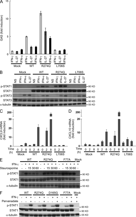
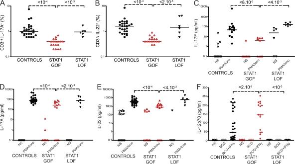
Chronic mucocutaneous candidiasis disease (CMCD) may be caused by autosomal dominant (AD) IL-17F deficiency or autosomal recessive (AR) IL-17RA deficiency. Here, using whole-exome sequencing, we identified heterozygous germline mutations in STAT1 in 47 patients from 20 kindreds with AD CMCD. Previously described heterozygous STAT1 mutant alleles are loss-of-function and cause AD predisposition to mycobacterial disease caused by impaired STAT1-dependent cellular responses to IFN-γ. Other loss-of-function STAT1 alleles cause AR predisposition to intracellular bacterial and viral diseases, caused by impaired STAT1-dependent responses to IFN-α/β, IFN-γ, IFN-λ, and IL-27. In contrast, the 12 AD CMCD-inducing STAT1 mutant alleles described here are gain-of-function and increase STAT1-dependent cellular responses to these cytokines, and to cytokines that predominantly activate STAT3, such as IL-6 and IL-21. All of these mutations affect the coiled-coil domain and impair the nuclear dephosphorylation of activated STAT1, accounting for their gain-of-function and dominance. Stronger cellular responses to the STAT1-dependent IL-17 inhibitors IFN-α/β, IFN-γ, and IL-27, and stronger STAT1 activation in response to the STAT3-dependent IL-17 inducers IL-6 and IL-21, hinder the development of T cells producing IL-17A, IL-17F, and IL-22. Gain-of-function STAT1 alleles therefore cause AD CMCD by impairing IL-17 immunity.


Figures

Figure 1.
Figure 1.
Heterozygous missense mutations affecting the STAT1 coiled-coil domain in kindreds with AD CMCD. (A) The human STAT1 α isoform is shown, with its known pathogenic mutations. Coding exons are numbered with roman numerals and delimited by a vertical bar. Regions corresponding to the coiled-coil domain (CC), DNA-binding domain (DNA-B), linker domain (L), SH2 domain (SH2), tail segment domain (TS), and transactivator domain (TA) are indicated, together with their amino-acid boundaries, and are delimited by bold lines. Tyr701 (pY) and Ser727 (pS) are indicated. Mutations in green are dominant and associated with partial STAT1 deficiency and MSMD. Mutations in brown are recessive and associated with complete STAT1 deficiency and intracellular bacterial and viral disease. Mutations in blue are recessive and associated with partial STAT1 deficiency and intracellular bacterial and/or viral disease. Mutations in red are dominant and associated with a gain-of-function of STAT1 and CMCD. (B) Pedigrees of 20 families with AD “gain-of-function” STAT1 mutations. Each kindred is designated by a letter (A to T), each generation is designated by a roman numeral (I-II-III-IV), and each individual is designated by an Arabic numeral (each individual studied is identified by a code of this type, organized from left to right). Black indicates CMCD patients. The probands are indicated by arrows. When tested, the genotype for STAT1 is indicated below each individual. (C) Three-dimensional structure of phosphorylated STAT1 in complex with DNA. Connolly surface representation, with the following amino acids highlighted: red, amino acids mutated in patients with CMCD; blue, amino acids located in the coiled-coil domain and mutated in patients with MSMD and viral diseases; yellow, amino acids identified in vitro as affecting the dephosphorylation process.
Figure 2.
Figure 2.
The mutant R274Q STAT1 allele is gain-of-phosphorylation and gain-of-function for GAF-dependent cellular responses. U3C cells were transfected with a mock vector, a WT, or two mutant alleles of STAT1 (R274Q and L706S). The response to IFN-γ, IL-27, and IFN-α was then evaluated by determining luciferase activity of a reporter gene under the control of the GAS promoter (A), and by determining STAT1 and STAT3 phosphorylation by Western blot (B). Experiments were performed at least three times independently. (C and D) Quantitative RT-PCR was used to measure the induction of CXCL9 (C) and CXCL10 (D) 2–8 h after stimulation with IFN-γ. Experiments were performed two times independently. (E) The nuclear dephosphorylation of STAT1 was tested by WB in U3C cells transfected with a mock vector, WT STAT1, the R274Q, or the F77A (a known loss-of-dephosphorylation mutant) STAT1 mutant alleles, and treated with IFN-γ with or without the tyrosine kinase inhibitor staurosporine for the indicated periods of time (in minutes). Three independent experiments were performed. (F) Western blot of U3C cells transfected with mock, WT, R274Q, D165G, and F77A alleles of STAT1, nontreated or treated with IFN-γ in the absence or presence of the phosphatase inhibitor pervanadate. Two independent experiments were performed. Error bars represent SD of one experiment done in triplicate (Fig. S1 D).
Figure 3.
Figure 3.
The mutant R274Q STAT1 allele is dominant for GAF-dependent cellular responses at the cellular level. The responses of the patient’s EBV-B cells (R274Q/WT) were evaluated independently at least twice, by EMSA, with a GAS probe (A, C, E, and G), and by Western blot (B, D, F, and H). This response was compared with that of one or two healthy controls (WT/WT1 and WT/WT2), heterozygous cells with a WT and a loss-of-function STAT1 allele (STAT1+/−), cells heterozygous for a dominant loss-of-function mutation of STAT1 (L706S/WT), cells with complete STAT1 deficiency (STAT1−/−), and cells from two patients heterozygous for dominant loss-of-function mutations of STAT3 (STAT3+/−1 and STAT3+/−2). Cells were left nonstimulated (NS) or stimulated, as indicated, with IFN-γ, IFN-α, IL-27, IL-6, and IL-21. pSTAT is an antibody specific for STAT with a phosphorylated tyrosine residue. (I) The nuclear and cytoplasmic fractions of EBV-B cells from a control (WT/WT), a CMCD patient (R274Q/WT), a heterozygous patient with a dominant loss-of-function mutation of STAT1 (L706S/WT) and a patient with complete STAT1 deficiency (−/−) stimulated with IFN-γ and IFN-α were tested for the presence of phosphorylated STAT1 and STAT1 by WB. Antibodies directed against GAPDH and Lamin B1 were used to normalize the amount of cytoplasmic and nuclear proteins, respectively. The experiment was performed twice.
Figure 4.
Figure 4.
Impaired development and function of IL-17– and IL-22–producing T cells ex vivo in patients with AD CMCD and STAT1 mutations. Each symbol represents a value from a healthy control individual (black circles), a patient bearing a STAT1 gain-of-function (GOF) allele (red upright triangles), or a patient bearing one or two STAT1 loss-of-function (LOF) alleles (black upside-down triangles). (A and B) Percentage of CD3+/IL-17A+ (A) and CD3+/IL-22+ (B) cells, as determined by flow cytometry, in nonadherent PBMCs activated by incubation for 12 h with PMA and ionomycin. (C–E) Secretion of IL-17F (C), IL-17A (D) and IL-22 (E) by whole blood cells, as determined by ELISA, in the absence of stimulation (open symbols) and after stimulation with PMA and ionomycin for 48 h (closed symbols). Horizontal bars represent medians. The p-values for the nonparametric Wilcoxon test, between patients with STAT1 GOF mutations (n = 18) and controls (n = 28) and patients with STAT1 LOF mutations (n = 6) are indicated. All differences between healthy controls and patients with STAT1 LOF alleles were not significant. (F) Secretion of IL-12p70 by whole blood cells, as determined by ELISA, in the absence of stimulation (open symbols), after stimulation with BCG (lightly colored symbols), or BCG + IFN-γ for 48 h (closed symbols). Horizontal bars represent medians. The p-values for differences between patients with STAT1 GOF mutations (n = 15) and controls (n = 23) and patients with STAT1 LOF mutations (n = 6) are indicated and were calculated in nonparametric Wilcoxon tests. All experiments were performed at least two times independently.

References

    1. Adzhubei I.A., Schmidt S., Peshkin L., Ramensky V.E., Gerasimova A., Bork P., Kondrashov A.S., Sunyaev S.R. 2010. A method and server for predicting damaging missense mutations. Nat. Methods. 7:248–249 10.1038/nmeth0410-248 - DOI - PMC - PubMed
    1. Alcaïs A., Abel L., Casanova J.L. 2009. Human genetics of infectious diseases: between proof of principle and paradigm. J. Clin. Invest. 119:2506–2514 10.1172/JCI38111 - DOI - PMC - PubMed
    1. Alcaïs A., Quintana-Murci L., Thaler D.S., Schurr E., Abel L., Casanova J.L. 2010. Life-threatening infectious diseases of childhood: single-gene inborn errors of immunity? Ann. N. Y. Acad. Sci. 1214:18–33 10.1111/j.1749-6632.2010.05834.x - DOI - PubMed
    1. Amadi-Obi A., Yu C.R., Liu X., Mahdi R.M., Clarke G.L., Nussenblatt R.B., Gery I., Lee Y.S., Egwuagu C.E. 2007. TH17 cells contribute to uveitis and scleritis and are expanded by IL-2 and inhibited by IL-27/STAT1. Nat. Med. 13:711–718 10.1038/nm1585 - DOI - PubMed
    1. Atkinson T.P., Schäffer A.A., Grimbacher B., Schroeder H.W., Jr, Woellner C., Zerbe C.S., Puck J.M. 2001. An immune defect causing dominant chronic mucocutaneous candidiasis and thyroid disease maps to chromosome 2p in a single family. Am. J. Hum. Genet. 69:791–803 10.1086/323611 - DOI - PMC - PubMed

Publication types

MeSH terms