. 2011 Aug 1;208(8):1661-71.
doi: 10.1084/jem.20101623. Epub 2011 Jul 4.

The immunoreceptor adapter protein DAP12 suppresses B lymphocyte-driven adaptive immune responses

Affiliations

The immunoreceptor adapter protein DAP12 suppresses B lymphocyte-driven adaptive immune responses

Takako Nakano-Yokomizo et al. J Exp Med. .

Abstract

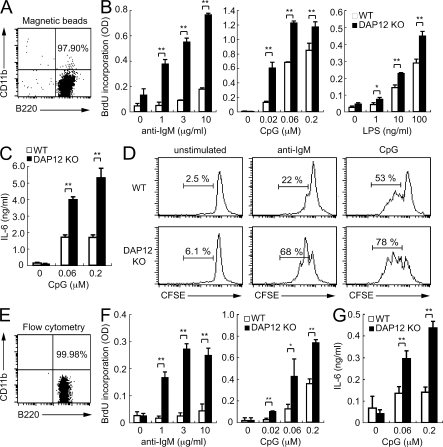
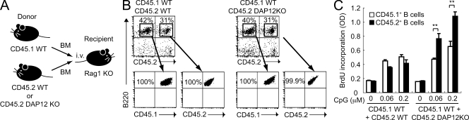
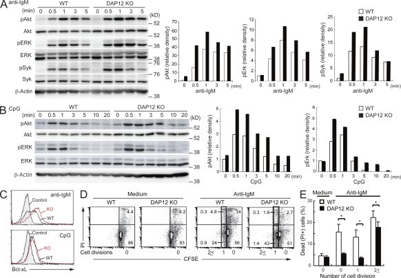
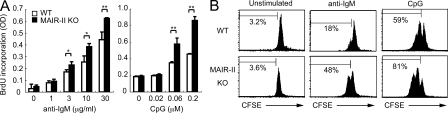
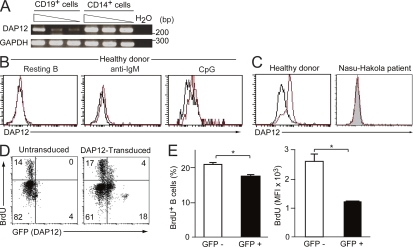
DAP12, an immunoreceptor tyrosine-based activation motif-bearing adapter protein, is involved in innate immunity mediated by natural killer cells and myeloid cells. We show that DAP12-deficient mouse B cells and B cells from a patient with Nasu-Hakola disease, a recessive genetic disorder resulting from loss of DAP12, showed enhanced proliferation after stimulation with anti-IgM or CpG. Myeloid-associated immunoglobulin-like receptor (MAIR) II (Cd300d) is a DAP12-associated immune receptor. Like DAP12-deficient B cells, MAIR-II-deficient B cells were hyperresponsive. Expression of a chimeric receptor composed of the MAIR-II extracellular domain directly coupled to DAP12 into the DAP12-deficient or MAIR-II-deficient B cells suppressed B cell receptor (BCR)-mediated proliferation. The chimeric MAIR-II-DAP12 receptor recruited the SH2 domain-containing protein tyrosine phosphatase 1 (SHP-1) after BCR stimulation. DAP12-deficient mice showed elevated serum antibodies against self-antigens and enhanced humoral immune responses against T cell-dependent and T cell-independent antigens. Thus, DAP12-coupled MAIR-II negatively regulates B cell-mediated adaptive immune responses.


Figures

Figure 1.
Figure 1.
Enhanced proliferation of DAP12-deficient B cells in response to anti-IgM and CpG. B cells were purified from spleens of WT or DAP12-deficient (DAP12 KO) mice by positive selection using magnetic beads coated with anti-B220 mAb (A), followed by cell sorting using flow cytometry (E), and were analyzed by flow cytometry. Representative data of the B cell purification from WT mice are shown (A and E). The purified B cells were stained or not with CFSE and stimulated with F(ab’)2 fragments of anti-IgM, CpG, or LPS. Proliferation was analyzed by BrdU incorporation (B and F) or flow cytometry (D). Anti-IgM at 3 µg/ml and CpG at 0.06 µM were used for stimulation in D. Culture supernatants were assayed for IL-6 production by ELISA (C and G). The proliferation assays shown in B, D, and F were done using B cells isolated by magnetic beads alone or by flow cytometry, respectively. *, P < 0.05; **, P < 0.01. Data are representative of more than three independent experiments. Error bars show SD.
Figure 2.
Figure 2.
Enhanced proliferation of DAP12-deficient B cells derived from BM chimeric mice in responses to CpG. (A) BM chimeric mice were generated by adoptive transfer of an equal number of BM cells isolated from CD45.1 WT mice and either CD45.2 WT or DAP12-deficient mice into Rag1-deficient mice. (B) CD45.1+ WT B cells and CD45.2+ WT or DAP12-deficient B cells were then sorted to >99.9% purity from spleens of mixed BM chimeric mice and were analyzed by flow cytometry. (C) The purified B cells were stimulated with CpG for 48 h. Proliferation was analyzed by BrdU incorporation. **, P < 0.01. Data are representative of three independent experiments. Error bars show SD.
Figure 3.
Figure 3.
Enhanced tyrosine phosphorylation of Erk, Akt, and Bcl-xL and prolonged survival in DAP12-deficient B cells. Purified B cells from the spleens of WT and DAP12-deficient mice were stimulated with 5 µg/ml of F(ab’)2 fragments of anti–mouse IgM (A and C) or 0.06 µM CpG (B and C). (A and B) The stimulated cells were lysed and then analyzed by immunoblotting with antibodies specific for indicated proteins. Bar graphs show the relative amount of each phosphorylated protein, as determined by densitometry, before and after stimulation. (C) The purified B cells 24 h after stimulation were fixed and stained with anti–Bcl-xL and then analyzed by flow cytometry. (D and E) The purified B cells were labeled with CFSE, stimulated with 10 µg/ml anti–mouse IgM or unstimulated (medium) for 48 h, stained with PI and analyzed by flow cytometry (D). The dead cell population was calculated as follows: PI+ cell frequency/PI+ and PI cell frequency in each division peak of CFSE dilution (E). *, P < 0.05. n = 4. Data are representative of five (A), three (B), two (C), and four (D) independent experiments. Error bars show SD.
Figure 4.
Figure 4.
Enhanced proliferation of MAIR-II–deficient B cells in response to anti-IgM and CpG. B cells were purified from the spleens of WT or MAIR-II–deficient mice, labeled with CFSE (B) or not (A), and stimulated with F(ab’)2 fragments of anti-IgM or CpG. Proliferation was analyzed by flow cytometry. *, P < 0.05; **, P < 0.01. Data are representative of three independent experiments.
Figure 5.
Figure 5.
DAP12-coupled MAIR-II inhibits B cell proliferation. (A) Schematic diagram of the constructs: GFP (control); WT DAP12 consisting of extracellular (EX), transmembrane (TM), and cytoplasmic (Cy) domains followed by GFP (DAP12); and the fusion protein consisting of the MAIR-II extracellular domain, followed by DAP12 containing an Ala (A) to Asp (D) substitution in the transmembrane domain (TM), followed by GFP (MAIR-II/DAP12). (B) Purified DAP12-deficient B cells were transduced with the lentiviral vectors and GFP expression was analyzed by flow cytometry. Shaded and open histograms indicate untransduced and transduced B cells, respectively. (C and D) The lentiviral vector–transduced DAP12-deficient or MAIR-II–deficient B cells were stimulated with PMA, F(ab’)2 fragments of anti-IgM, or CpG and then analyzed for proliferation. Error bars show SD. (E) K46 µmλ cells were transduced with the MAIR-II/DAP12 lentiviral vector, stained with anti–MAIR-II or anti-IgM, and analyzed by flow cytometry (left). K46 µmλ transfectants were stimulated with anti-IgM, and immunoprecipitates with anti–MAIR-II or control Ig were immunoblotted with anti-SHIP, anti–SHP-1, or anti–MAIR-II (right). *, P < 0.05; ***, P < 0.001. Data are representative of three independent experiments.
Figure 6.
Figure 6.
Enhanced humoral immune responses in DAP12-deficient mice. (A–H) WT (n ≈ 5–9) and DAP12-deficient (n ≈ 5–9) mice were depleted (F–H) or not (A–E) of CD4+ T cells and macrophages by injection with anti-CD4 mAb and Cl2MBP liposomes, as described in Fig. S5, and then immunized with NP-CGG (A–C) or NP-Ficoll (D–H). 14 (A–C) or 8 (D–H) d after the immunization, spleen sections were stained with biotinylated PNA, followed by Alexa Fluor 594–conjugated streptavidin in combination with FITC-conjugated anti-B220 (A, D, and F). Spleen cells were also stained with FITC-conjugated GL7 mAb, biotin-conjugated PNA, and APC-conjugated anti-B220, followed by PE-conjugated streptavidin, and the frequencies of GC B cells (defined as the percentage of GL-7+PNA+B220+ cells) were determined by flow cytometry (B, E, and G). Sera were collected from these mice and analyzed for NP-specific antibodies by ELISA (C and H). (I and J) Mixed BM chimeric mice were generated by adoptive transfer of an equal number of BM cells isolated from CD45.1 WT mice and CD45.2 DAP12-deficient mice into Rag1-deficient mice. The spleen sections from the BM chimeric mice 8 d after immunization with NP-Ficoll were analyzed by immunohistology, as described (I). The spleen cells from the same mice after the immunization were stained with anti-B220, GL7, biotinylated PNA, and either anti-CD45.1 or anti-CD45.2, followed by APC-conjugated streptavidin. The proportions of CD45.1 WT or CD45.2 DAP12-deficient B cells in non-GC (GL7PNAB220+) or GC (GL7+PNA+B220+) B cells are indicated (J). (K) Sera were collected from WT (n = 5) and DAP12-deficient (n = 5) mice at the age of 10 wk old and analyzed for the titer of antibodies specific for the self-antigens indicated. The horizontal bars show the mean. *, P < 0.05; **, P < 0.01; ***, P < 0.001. Data are representative of five mice (A, D, and F) and two (B, E, G, I, and J) and three (K) independent experiments. Error bars show SD.
Figure 7.
Figure 7.
DAP12 inhibits human B cell proliferation. (A) PBMCs derived from a healthy donor were stained with PE-conjugated anti-CD19 and FITC-conjugated anti-CD14 mAbs. CD19+ B cells or CD14+ monocytes were sorted to >99.9% purity by flow cytometry. The RNA was extracted from the purified cells and was subjected to semiquantitative RT-PCR for expression of DAP12 and GAPDH transcripts, according to template dose by dilution. (B and C) PBMCs derived from a healthy donor or a patient with Nasu-Hakola disease were stimulated (B) or not (C) with anti-IgM or CpG for 48 h and then stained with PE-conjugated anti-CD20 (B) or anti-CD14 (C). Cells were fixed and then stained with anti-DAP12 (red line) or control Ig (black line), followed by FITC-conjugated secondary antibody. Plots in B are gated on CD20+ cells and plots in C are gated on CD14+ cells. (D and E) PBMCs from a patient with Nasu-Hakola disease were transduced or not with the lentiviral vector encoding DAP12 and EGFP and then stimulated with CpG. BrdU was pulsed for the final 24 h in the culture. Cells were stained with PE-conjugated anti-CD19 mAb and then fixed, followed by staining with APC-conjugated anti-BrdU mAb and analyzed for B cell proliferation, as determined by BrdU incorporation, on the CD19+ gated cells by flow cytometry (D). The frequencies of BrdU+ cells in GFP and GFP+ cells and mean fluorescence intensity (MFI) of BrdU+ cells in GFP and GFP+ cells were determined (E). The data represent two independent experiments (A–E). *, P < 0.05. Error bars show SD.

References

    1. Allman D., Lindsley R.C., DeMuth W., Rudd K., Shinton S.A., Hardy R.R. 2001. Resolution of three nonproliferative immature splenic B cell subsets reveals multiple selection points during peripheral B cell maturation. J. Immunol. 167:6834–6840 - PubMed
    1. Bakker A.B., Hoek R.M., Cerwenka A., Blom B., Lucian L., McNeil T., Murray R., Phillips L.H., Sedgwick J.D., Lanier L.L. 2000. DAP12-deficient mice fail to develop autoimmunity due to impaired antigen priming. Immunity. 13:345–353 10.1016/S1074-7613(00)00034-0 - DOI - PubMed
    1. Barrow A.D., Trowsdale J. 2006. You say ITAM and I say ITIM, let’s call the whole thing off: the ambiguity of immunoreceptor signalling. Eur. J. Immunol. 36:1646–1653 10.1002/eji.200636195 - DOI - PubMed
    1. Blank U., Launay P., Benhamou M., Monteiro R.C. 2009. Inhibitory ITAMs as novel regulators of immunity. Immunol. Rev. 232:59–71 10.1111/j.1600-065X.2009.00832.x - DOI - PubMed
    1. Blasius A.L., Cella M., Maldonado J., Takai T., Colonna M. 2006. Siglec-H is an IPC-specific receptor that modulates type I IFN secretion through DAP12. Blood. 107:2474–2476 10.1182/blood-2005-09-3746 - DOI - PMC - PubMed

Publication types

MeSH terms

Substances

Supplementary concepts