

Is pocket mobile echocardiography the next-generation stethoscope? A cross-sectional comparison of rapidly acquired images with standard transthoracic echocardiography
- PMID: 21727291
- PMCID: PMC3733444
- DOI: 10.7326/0003-4819-155-1-201107050-00005
Is pocket mobile echocardiography the next-generation stethoscope? A cross-sectional comparison of rapidly acquired images with standard transthoracic echocardiography
Abstract
Background: A pocket mobile echocardiography (PME) device is commercially available for clinical use, but public data documenting its accuracy compared with standard transthoracic echocardiography (TTE) are not available.
Objective: To compare the accuracy of rapidly acquired PME images with those acquired by standard TTE.
Design: Cross-sectional study. At the time of referral for TTE, ultrasonographers acquired PME images first in 5 minutes or less. Ultrasonographers were not blinded to the clinical indication for imaging or to the PME image results when obtaining standard TTE images. Two experienced echocardiographers and 2 cardiology fellows who were blinded to the indication for the study and TTE results but not to the device source interpreted the PME images.
Setting: Scripps Clinic Torrey Pines and Scripps Green Hospital, La Jolla, California.
Patients: Convenience sample of 97 patients consecutively referred for echocardiography.
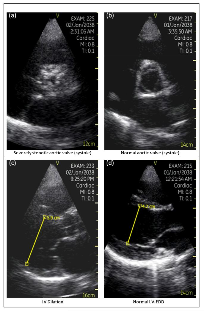
Measurements: Visualizability and accuracy (the sum of proportions of true-positive and true-negative readings and observer variability) for ejection fraction, wall-motion abnormalities, left ventricular end-diastolic dimension, inferior vena cava size, aortic and mitral valve pathology, and pericardial effusion.
Results: Physician-readers could visualize some but not all echocardiographic measurements obtained with the PME device in every patient (highest proportions were for ejection fraction and left ventricular end-diastolic dimension [95% each]; the lowest proportion was for inferior vena cava size [75%]). Accuracy also varied by measurement (aortic valve was 96% [highest] and inferior vena cava size was 78% [lowest]) and decreased when nonvisualizability was accounted for (aortic valve was 91% and inferior vena cava size was 58%). Observer agreement was fair to moderate for some measurements among less-experienced readers.
Limitation: The study was conducted at a single setting, there was no formal estimate of accuracy given the small convenience sample of patients, and few abnormal echocardiographic measurements occurred.
Conclusion: The rapid acquisition of images by skilled ultrasonographers who use PME yields accurate assessments of ejection fraction and some but not all cardiac structures in many patients. Further testing of the device in larger patient cohorts with diverse cardiac abnormalities and with untrained clinicians obtaining and interpreting images is required before wide dissemination of its use can be recommended.
Primary funding source: National Institutes of Health.
Figures

References
-
- Kimura BJ, Demaria AN. Empowering physical examination: the “laying on” of ultrasound. JACC Cardiovasc Imaging. 2008;1(5):602–604. - PubMed
-
- Kobal SL, Atar S, Siegel RJ. Hand-carried ultrasound improves bedside cardiovascular examination. Chest. 2004;126(3):693–701. - PubMed
-
- Brennan JM, Blair JE, Goonewardena S, Ronan A, Shah D, Vasaiwala S, Kirkpatrick JN, Spencer KT. Reappraisal of the use of inferior vena cava for estimating right atrial pressure. J Am Soc Echocardiogr. 2007;20(7):857–861. - PubMed
-
- Popp RL. The physical exam of the future: echocardiography as part of the assessment. ACC Curr. J. Rev. 1998;7:79–81.
Publication types
MeSH terms
Grants and funding
LinkOut - more resources
Full Text Sources
Research Materials