. 2011 Jul 5:4:127.
doi: 10.1186/1756-3305-4-127.

Fibrinogen-related proteins in ixodid ticks

Affiliations

Fibrinogen-related proteins in ixodid ticks

Jan Sterba et al. Parasit Vectors. .

Abstract

Background: Fibrinogen-related proteins with lectin activity are believed to be part of the tick innate immune system. Several fibrinogen-related proteins have been described and characterised mainly on the basis of their cDNA sequences while direct biochemical evidence is missing. One of them, the haemolymph lectin Dorin M from the tick Ornithodoros moubata was isolated and characterised in more depth.

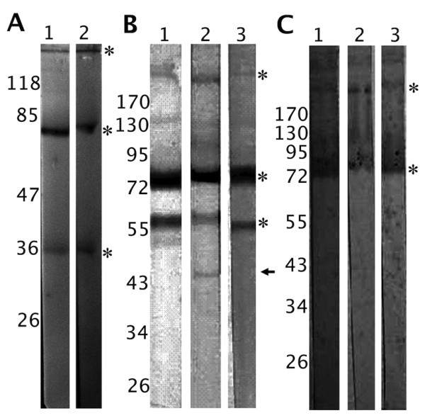
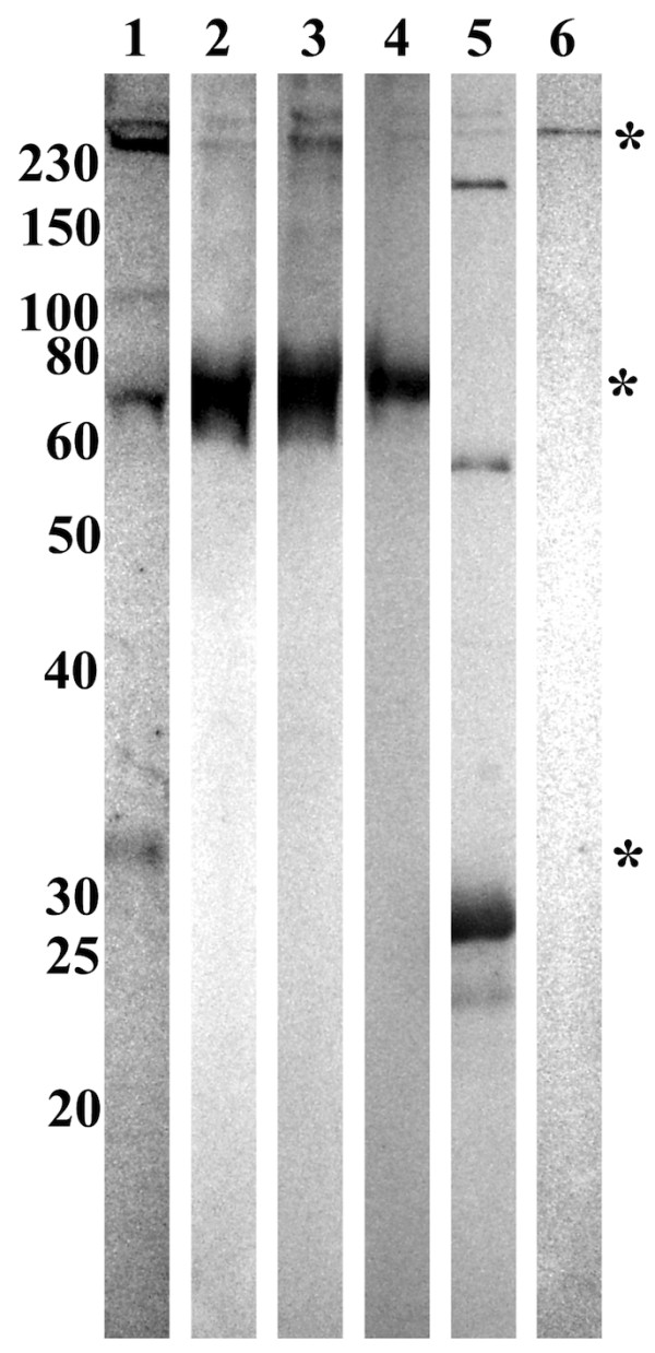
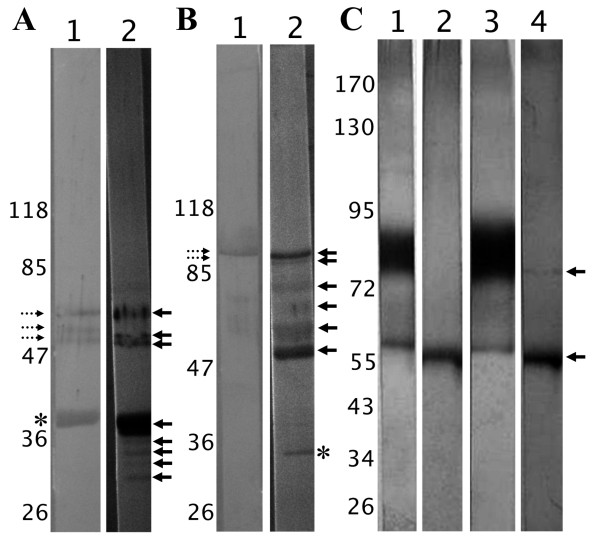
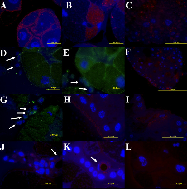
Results: Several fibrinogen-related proteins were detected in the haemolymph of ixodid ticks Dermacentor marginatus, Rhipicephalus appendiculatus, R. pulchellus, and R. sanguineus. These proteins were recognised by sera directed against the tick lectin Dorin M and the haemagglutination activity of the ticks R. appendiculatus and D. marginatus. Cross-reactivity of the identified proteins with antibodies against the fibrinogen domain of the human ficolin was also shown. The carbohydrate-binding ability of tick haemolymph was confirmed by haemagglutination activity assays, and this activity was shown to be inhibited by neuraminic acid and sialylated glycoproteins as well as by N-acetylated hexosamines. The fibrinogen-related proteins were shown to be glycosylated and they were localised in salivary glands, midguts, and haemocytes of D. marginatus. Hemelipoglycoprotein was also recognised by sera directed against the fibrinogen-related proteins in all three Rhipicephalus species as well as in D. marginatus. However, this protein does not contain the fibrinogen domain and thus, the binding possibly results from the structure similarity between hemelipoglycoprotein and the fibrinogen domain.

Conclusions: The presence of fibrinogen-related proteins was shown in the haemolymph of four tick species in high abundance. Reactivity of antibodies directed against ficolin or fibrinogen-related proteins with proteins which do not contain the fibrinogen domain points out the importance of sequence analysis of the identified proteins in further studies. Previously observed expression of fibrinogen-related proteins in haemocytes together with the results of this study suggest involvement of fibrinogen-related proteins in tick immunity processes. Thus, they have potential as targets for anti-tick vaccines and as antimicrobial proteins in pharmacology. Research on fibrinogen-related proteins could reveal further details of tick innate immunity processes.


Figures

Figure 1
Figure 1
Immunodetection of the putative fibrinogen-related proteins in tick haemolymph. 1A - Electrophoretically separated and electroblotted non-reduced D. marginatus haemolymph proteins immunostained using mouse anti-(DM)HA serum (lane 1) and rabbit anti-Dorin M serum (lane 2). Four proteins were detected with molecular weights of approximately 36, 79/80, and 290 kDa (marked with asterisks; the 79/80 kDa double-band is marked by one asterisk). 1B - Electrophoretically separated and electroblotted non-reduced R. appendiculatus (lane 1), R. pulchellus (lane 2), and R. sanguineus (lane 3) haemolymph proteins immunostained using mouse anti-(RA)HA serum. In each sample, three proteins were detected with size of 58, 75, and 185 kDa (asterisks). Furthermore, a protein band with molecular weight of 45 kDa was observed in R. pulchellus (arrow). 1C - Electrophoretically separated and electroblotted non-reduced R. appendiculatus (lane 1), R. pulchellus (lane 2), and R. sanguineus (lane 3) haemolymph proteins immunostained using rabbit anti-Dorin M serum. Proteins with molecular weight of 75 and 185 kDa were detected, as in the case of anti-(RA)HA serum (asterisks).
Figure 2
Figure 2
Immunoblotting of tick haemolymph proteins with anti-ficolin antibodies. Electrophoretically separated and electroblotted non-reduced haemolymph proteins from D. marginatus (lane 1), R. appendiculatus (lane 2), R. pulchellus (lane 3), and R. sanguineus (lane 4) were immunostained using rabbit anti-FCN1 H antibodies. Recombinant human ficolin 1 was used as a control (lane 5). Purified hemelipoglycoprotein from D. marginatus haemolymph, which was identified by MS as one of the recognised proteins was used as a control (lane 6). However, this protein does not contain the fibrinogen domain [3]. The same proteins as in Figure 1A were detected in D. marginatus haemolymph (36 kDa, 79/80 kDa, and 290 kDa proteins; marked with asterisks; the 79/80 kDa double-band is marked by one asterisk). In Rhipicephalus ticks haemolymphs, the 72 kDa and 290 kDa proteins were detected, but not the 55 kDa protein. Additionally, the purified hemelipoglycoprotein from D. marginatus was detected by the anti-FCN1 H antibodies. Non-reduced recombinant human ficolin 1 served as a positive control. Antibodies positively reacted with subunits of the protein (approximately 30 kDa) as well as with higher molecular weight complexes (approximately 60 kDa, 180 kDa, 250 kDa, 280 kDa).
Figure 3
Figure 3
Deglycosylation of the putative fibrinogen-related proteins in tick haemolymph and their immunodetection. 3A - Reduced D. marginatus haemolymph proteins (lane 1) were enzymatically deglycosylated (lane 2). The FReP proteins were detected using anti-DMF1 serum, which was raised against the 36 kDa protein (asterisk). After deglycosylation, additional bands appeared with sizes of 31, 33, and 34 kDa (arrows). Cross-reactivity of the serum with 79/80 kDa proteins was observed (bands with molecular weights of 58, 60, and 66 kDa - lane 1, dotted arrows). The molecular weight of these proteins shifted after deglycosylation and three bands were observed at 54, 58, and 63 kDa (lane 2, arrows). 3B - Reduced D. marginatus haemolymph proteins (lane 1) a were enzymatically deglycosylated (lane 2). The FReP proteins were detected using anti-DMF3 serum, which was raised against the 290 kDa protein. This protein is composed of two subunits which have 95 and 100 kDa (lane 1, dotted arrows). After deglycosylation (lane 2), additional bands appeared at 50 and 74 kDa (arrows). We observed cross-reactivity of the serum also in this case, when the same bands were observed for the 79/80 kDa proteins (lane 2, arrows) as in the case of anti-DMF1 serum (see Figure 2A). The protein band at 34 kDa (lane 2, asterisk) is probably a protein cleavage product. 3C - Reduced R. appendiculatus (lanes 1,2) and R. sanguineus (lanes 3,4) haemolymph proteins before (lanes 1,3) and after deglycosylattion (lanes 2,4). The FReP proteins were detected using anti-(RA)HA serum. In both tick samples (lanes 1,3), 58 kDa band was observed and a protein smear from 75 to 90 kDa (75 kDa protein and the subunits for 185 kDa protein). The deglycosylation diminished the reactivity of the protein smear and only the band at 58 kDa remained visible (lanes 2,4; arrows).
Figure 4
Figure 4
Immunolocalisation of FRePs in D. marginatus organs using fluorescence microscopy. Thin sections of midgut (C, F, H, I), salivary glands (A, B, J, K, L), and haemocytes attached to salivary glands (D, E, G) from fed female D. marginatus were labelled with sera raised against D. marginatus FRePs. Anti-DMF1 (A, B, C, D, E), anti-DMF2 (F, G), and anti-DMF3 (H, I, J, K) antibodies were used. Control reaction were carried out, in which anti-DMF sera were omitted (L). FRePs were localised on the surface and inside epithelial cells surrounding secretory granular cells of the acini type II (A, D, E); in granules of secretory cells located in the acini type III (B) and in haemocytes attached to the surface of salivary glands (D, E, G; arrows). Positive labelling reactions were observed in the cytoplasm of epithelium of salivary duct cells (J, K; arrows) and in thin cuticular layer facing to the lumen of the salivary duct (J, K). Anti-DMF sera detected structures on the surface (H) and inside the midgut cells (C, F, I). A, B, C, F, H, I, J, K, and L - Cy3-conjugated secondary antibodies (red); D, E, and G - FITC conjugated secondary antibodies (green). Cell nuclei were counter-stained using DAPI (blue).

References

    1. Sonenshine DE. Biology of ticks. Vol. 2. New York: Oxford University Press; 1993.
    1. Piesman J, Gage KL. In: The biology of disease vectors. Beaty BJ, Marquardt WC, editor. Niwot, CO: University of Colorado Press; 1996. Ticks and mites and the agents they transmit; pp. 160–174.
    1. Donohue KV, Khalil SMS, Mitchell RD, Sonenshine DE, Roe RM. Molecular characterization of the major hemelipoglycoprotein in ixodid ticks. Insect Mol Biol. 2008;17(3):197–208. doi: 10.1111/j.1365-2583.2008.00794.x. - DOI - PubMed
    1. Dupejova J, Sterba J, Vancova M, Grubhoffer L. Hemelipoglycoprotein from the ornate sheep tick, Dermacentor marginatus: structural and functional characterization. Parasit Vectors. 2011;4:4. doi: 10.1186/1756-3305-4-4. - DOI - PMC - PubMed
    1. Johns R, Sonenshine DE, Hynes WL. Identification of a defensin from the hemolymph of the American dog tick, Dermacentor variabilis. Insect Biochem Molec. 2001;31(9):857–865. doi: 10.1016/S0965-1748(01)00031-5. - DOI - PubMed

Publication types

MeSH terms

LinkOut - more resources