. 2011 Jul 6:9:47.
doi: 10.1186/1741-7007-9-47.

Structural and functional basis for RNA cleavage by Ire1

Affiliations

Structural and functional basis for RNA cleavage by Ire1

Alexei V Korennykh et al. BMC Biol. .

Abstract

Background: The unfolded protein response (UPR) controls the protein folding capacity of the endoplasmic reticulum (ER). Central to this signaling pathway is the ER-resident bifunctional transmembrane kinase/endoribonuclease Ire1. The endoribonuclease (RNase) domain of Ire1 initiates a non-conventional mRNA splicing reaction, leading to the production of a transcription factor that controls UPR target genes. The mRNA splicing reaction is an obligatory step of Ire1 signaling, yet its mechanism has remained poorly understood due to the absence of substrate-bound crystal structures of Ire1, the lack of structural similarity between Ire1 and other RNases, and a scarcity of quantitative enzymological data. Here, we experimentally define the active site of Ire1 RNase and quantitatively evaluate the contribution of the key active site residues to catalysis.

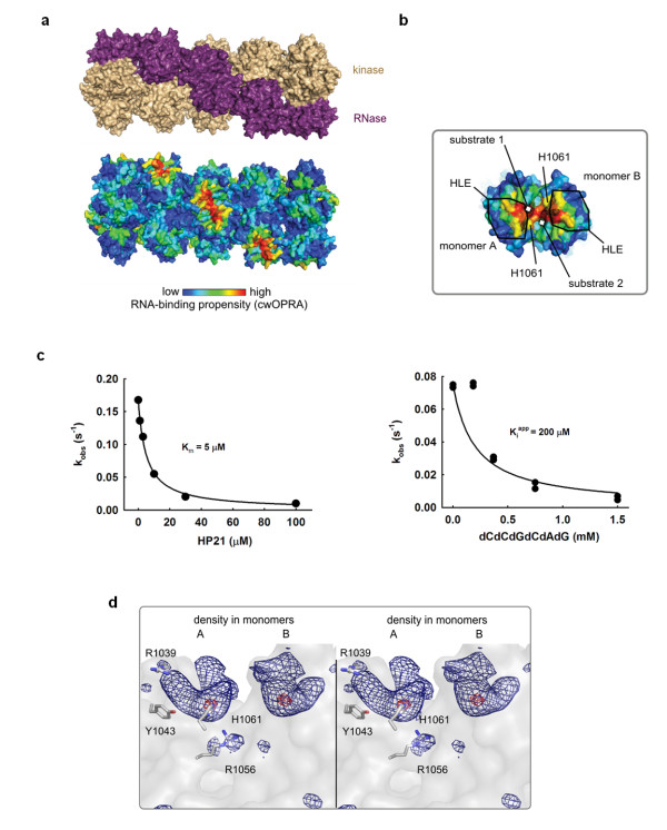
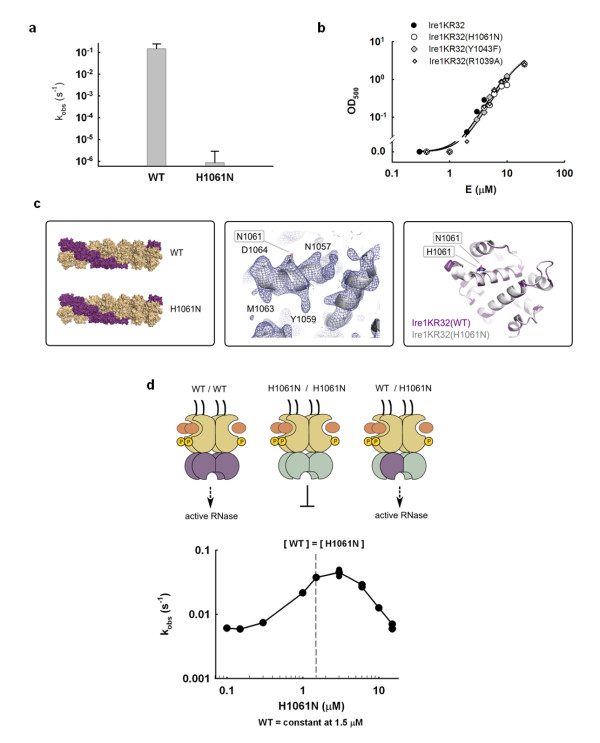
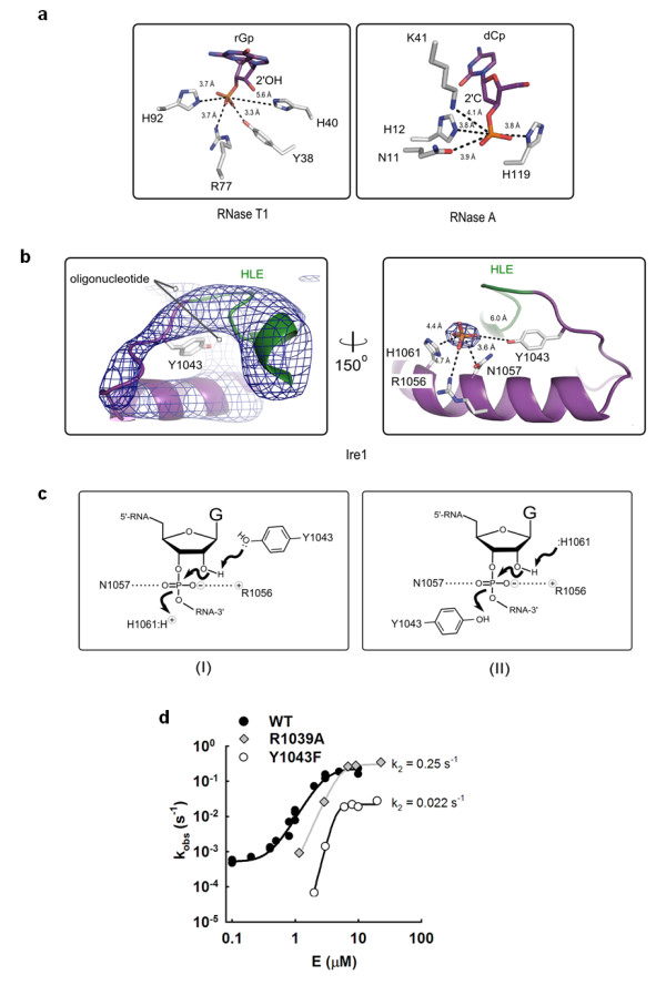
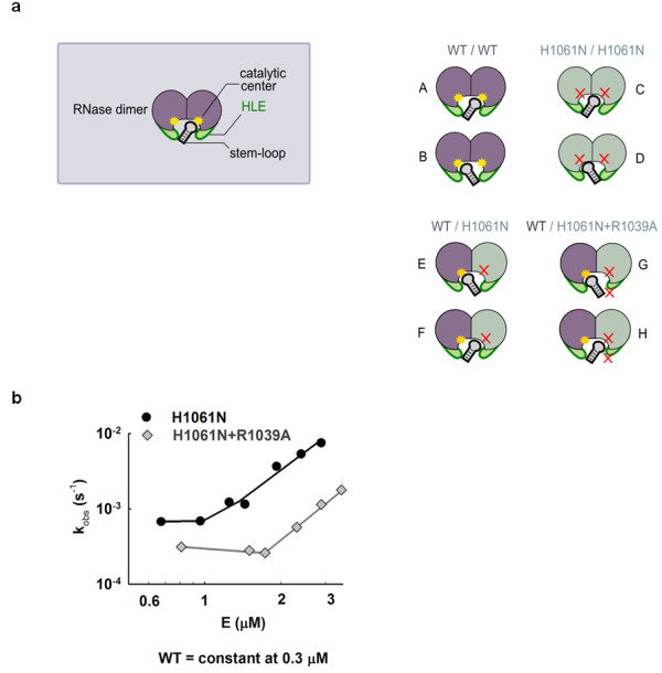
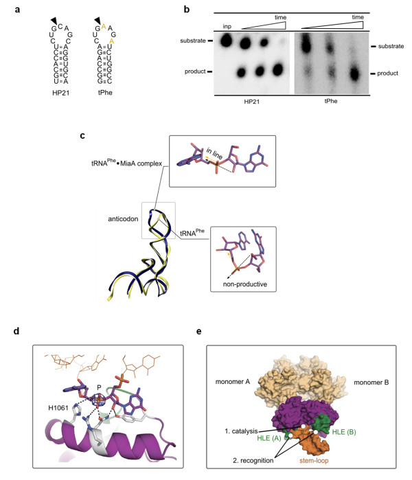
Results: This analysis and two new crystal structures suggest that Ire1 RNase uses histidine H1061 and tyrosine Y1043 as the general acid-general base pair contributing ≥7.6 kcal/mol and 1.4 kcal/mol to transition state stabilization, respectively, and asparagine N1057 and arginine R1056 for coordination of the scissile phosphate. Investigation of the stem-loop recognition revealed that additionally to the stem-loops derived from the classic Ire1 substrates HAC1 and Xbp1 mRNA, Ire1 can site-specifically and rapidly cleave anticodon stem-loop (ASL) of unmodified tRNAPhe, extending known substrate specificity of Ire1 RNase.

Conclusions: Our data define the catalytic center of Ire1 RNase and suggest a mechanism of RNA cleavage: each RNase monomer apparently contains a separate catalytic apparatus for RNA cleavage, whereas two RNase subunits contribute to RNA stem-loop docking. Conservation of the key residues among Ire1 homologues suggests that the mechanism elucidated here for yeast Ire1 applies to Ire1 in metazoan cells, and to the only known Ire1 homologue RNase L.


Figures

Figure 1
Figure 1
Oligonucleotide binding to Ire1 RNase. (a) Ire1 oligomer (PDB ID 3fbv) colored by domains (top) and RNA-binding propensity (bottom). (b) Close view of symmetric back-to-back RNase dimer from the oligomer in (a) with electron density for dCdCdGdCdAdG from the C222 crystal structure superimposed. Electron density colocalizes with the area of the strongest RNA binding propensity and is in direct contact with conserved histidine H1061 and the helix-loop element (HLE). (c) Binding of an RNA stem-loop HP21 and of dCdCdGdCdAdG to Ire1KR32 measured via inhibition of 32P-5'-HP21 cleavage by unlabeled HP21 (Km = 5 ± 1 μM) or dCdCdGdCdAdG (Kiapp = 200 ± 50 μM). Reactions contained 20 mM 4-(2-hydroxyethyl)-1-piperazine-ethanesulfonic acid (HEPES) (pH 7.4), 70 mM NaCl, 5% glycerol, 2 mM Mg(CH3COO)2, 2 mM ADP, 4 mM dithiothreitol, 3 μM total Ire1KR32 and ≤ 1 pM 32P-5'-HP21, and were conducted at 30°C. Measurements of HP21 binding at 1 μM and 10 μM of total Ire1KR32 are shown in Additional file 1, Figure S1. (d), Wall-eyed stereoview of non-crystallographic symmetry (NCS)-averaged Fo-Fc electron density map at the RNase active site. Contour levels are 7σ (dark blue) and 13σ (red). Side chain positions are from the crystal structure 3fbv.
Figure 2
Figure 2
Probing the role of H1061 in catalysis. (a) Effect of H1061N mutation on the RNase activity of Ire1. Error bars show standard error of single-exponential fitting. (b) Oligomerization profiles of Ire1KR32 and three mutants, Ire1KR32(H1061N), Ire1KR32(Y1043F) and Ire1KR32(R1039A) measured using light absorbance at 500 nM (see Methods). Measurements were conducted at room temperature using the same reaction buffers as in Figure 1c. (c) Comparison of Ire1 structures from oligomers formed by wild-type Ire1 (PDB ID 3fbv, space group P21212) and by H1061N mutant (new 3.65Å crystal structure, also in space group P21212). Shown are overall oligomer architectures (left), 2Fo-Fc electron density map for the H1061N RNase domain contoured at 1.3σ (middle) and superposition of the RNase domains (right). Simulated annealing Fo-Fc omit map for H1061N calculated without non-crystallographic symmetry (NCS) is shown in Additional file 1, Figure S2. (d) In trans activation of Ire1KR32 by the non-catalytic mutant H1061N. Concentration of wild-type Ire1KR32 was 1.5 μM, concentration of the H1061N Ire1 varied between 0.1-15 μM. Arrow marks the point of equivalent concentrations. Reactions were conducted under single-turnover conditions using the same reaction buffer as in Figure 1c.
Figure 3
Figure 3
Catalytic mechanism of Ire1 RNase. (a) Catalytic residues of RNase T1 and RNase A with productively bound nucleotides (PDB ID 1r5c and 1rga). (b) Left panel: simulated annealing (1000 K) omit-electron density map Fo-Fc for the helix-loop element (HLE; green) and adjacent loop containing Y1043 and the helix α4 of RNase domain (contoured at 5σ). Refinement was conducted with residues 1032-1057 deleted from all 7 monomers in the asymmetric unit, without using non-crystallographic symmetry (NCS). An alternative view of the electron density (after rotation around vertical axis) is shown in Additional file 1, Figure S4. Right panel: position of the scissile phosphate in the active site of Ire1 RNase. The strongest peak of the electron density (Fo-Fc, contoured at 12σ) was used to position the phosphate. Side chains were taken from PDB ID 3fbv). (c) Two plausible catalytic mechanisms in the active site of Ire1 RNase. (d) Enzyme titration profiles for Ire1KR32, Ire1KR32(Y1043F) and for Ire1KR32(R1039A) under single-turnover conditions. Reactions were conducted as in Figure 1c.
Figure 4
Figure 4
In trans activation of wild-type Ire1 RNase by Ire1 with RNase with a double mutation. (a) Schematic representation of RNase dimers formed via interface IF1c [5]. Due to a twofold symmetry, each RNase dimer can accommodate RNA stem-loop in two equivalent orientations (such as A and B). Both orientations are productive for wild-type Ire1. Neither orientation is productive for the H1061N mutant (C) and (D) due to disrupted catalysis. Only one orientation (E) is productive for the wild-type/H1061N chimera. Only one orientation (G) is also productive for the wild-type/H1061N+R1039A chimera, however this orientation is impaired by the R1039A mutation in the helix-loop element (HLE). (b) Titration of wild-type Ire1KR32 with Ire1KR32(H1061N) single mutant and Ire1KR32(H1061N, R1039A) double mutant. Reactions were conducted as in Figure 2d but contained 0.3 μM wild-type Ire1KR32 throughout the titrations to ensure subsaturating (k2/K1/2) regime for RNA cleavage.
Figure 5
Figure 5
Model of stem-loop recognition via a composite protein surface. (a) Stem-loops derived from XBP1 mRNA (HP21) and phenylalanine tRNAPhe (tPhe). Loop sequence differences are colored yellow in tPhe. (b) Time courses (0-5 min) for cleavage of 5'-32P-labeled stem-loops HP21 and tPhe by Ire1KR32 (3 μM) analyzed by polyacrylamide gel electrophoresis. (c) Superposition of crystal structures of tRNAPhe (PDB ID 1ehz) and tRNAPhe from tRNAPhe·MiaA complex (PDB ID 2zm5). Black arrow shows direction of nucleophile attack, orange arrow shows direction of leaving group. (d) Manual rigid-body docking of the base-flipped tRNAPhe anticodon into Ire1 crystal structure. Electron density for the scissile phosphate (contoured at 12σ) and the proposed catalytic mechanism (Figure 3c-I) were used as docking constraints. P designates scissile phosphate. The helix-loop element (HLE) is colored green. (e) Surface rendering of the model in (d). The model suggests cleavage of a stem-loop by one active site and recognition by a composite RNA binding surface.

References

    1. Cox JS, Walter P. A novel mechanism for regulating activity of a transcription factor that controls the unfolded protein response. Cell. 1996;87:391–404. doi: 10.1016/S0092-8674(00)81360-4. - DOI - PubMed
    1. Shamu CE, Walter P. Oligomerization and phosphorylation of the Ire1p kinase during intracellular signaling from the endoplasmic reticulum to the nucleus. EMBO J. 1996;15:3028–3039. - PMC - PubMed
    1. Sidrauski C, Cox JS, Walter P. tRNA ligase is required for regulated mRNA splicing in the unfolded protein response. Cell. 1996;87:405–413. doi: 10.1016/S0092-8674(00)81361-6. - DOI - PubMed
    1. Mori K, Ma W, Gething MJ, Sambrook J. A transmembrane protein with a cdc2+/CDC28-related kinase activity is required for signaling from the ER to the nucleus. Cell. 1993;74:743–756. doi: 10.1016/0092-8674(93)90521-Q. - DOI - PubMed
    1. Korennykh AV, Egea PF, Korostelev AA, Finer-Moore J, Zhang C, Shokat KM, Stroud RM, Walter P. The unfolded protein response signals through high-order assembly of Ire1. Nature. 2009;457:687–693. doi: 10.1038/nature07661. - DOI - PMC - PubMed

Publication types

MeSH terms

Substances