Review
. 2011 Aug;132(8-9):405-11.
doi: 10.1016/j.mad.2011.06.005. Epub 2011 Jun 25.

The excitatory neurotransmitter glutamate stimulates DNA repair to increase neuronal resiliency

Affiliations
Review

The excitatory neurotransmitter glutamate stimulates DNA repair to increase neuronal resiliency

Jenq-Lin Yang et al. Mech Ageing Dev. 2011 Aug.

Abstract

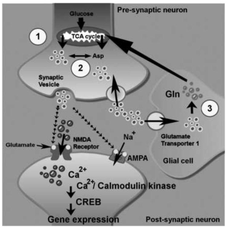
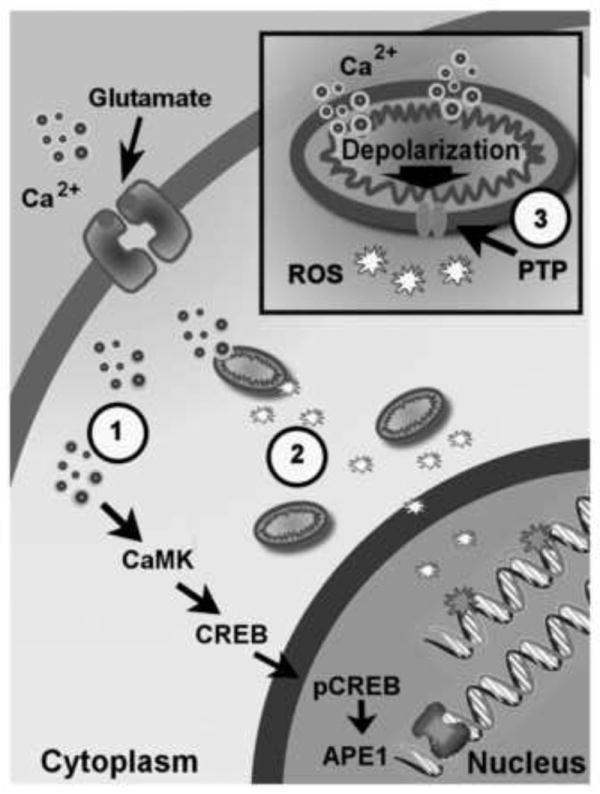
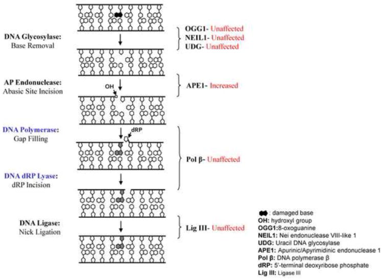
Glutamate is the most abundant excitatory neurotransmitter in the vertebrate central nervous system and plays an important role in synaptic plasticity required for learning and memory. Activation of glutamate ionotropic receptors promptly triggers membrane depolarization and Ca(2+) influx, resulting in the activation of several different protein kinases and transcription factors. For example, glutamate-mediated Ca(2+) influx activates Ca(2+)/calmodulin-dependent kinase, protein kinase C, and mitogen activated protein kinases resulting in activation of transcription factors such as cyclic AMP response element binding protein (CREB). Abnormally prolonged exposure to glutamate causes neuronal injury, and such "excitotoxicity" has been implicated in many acute and chronic diseases including ischemic stroke, epilepsy, amyotrophic lateral sclerosis, Alzheimer's, Huntington's and Parkinson's diseases. Interestingly, although glutamate-induced Ca(2+) influx can cause DNA damage by a mitochondrial reactive oxygen species-mediated mechanism, the Ca(2+) simultaneously activates CREB, resulting in up-regulation of the DNA repair and redox protein apurinic/apyrimidinic endonuclease 1. Here, we review connections between physiological or aberrant glutamate receptor activation, Ca(2+)-mediated signaling, oxidative DNA damage and repair efficiency, and neuronal vulnerability. We conclude that glutamate signaling involves an adaptive cellular stress response pathway that enhances DNA repair capability, thereby protecting neurons against injury and disease.


Figures

Figure 1
Figure 1
Glutamate synthesis, recycling, and signaling in neuronal cells. The glutamate concentration is well maintained in central nervous system (CNS). Glutamate (light ringed circles) is commonly derived from products of TCA (citric acid cycle) (circle number 1) and converts from Aspartate (Asp) by aspartate transaminase (circle number 2). The released glutamate in the synaptic gap can be promptly sequestered into neurons and/or glial cells via glutamate transporter 1. The recycled glutamate is converted into glutamine by glutaminase in glial cells. Then, glutamine is delivered to neurons and converted into glutamate by glutamate synthetase (circle number 3). The activation of α-amino-3-hydroxy-5-methyl-4-isoxazolepropionic acid receptor (AMPA) receptors by glutamate binding trigger Na+ influx and consequently cause membrane depolarization. In the meantime, the depolarized membrane facilitates the Ca2+ influx (dark ringed circles) from activated N-Methyl-d-aspartic acid (NMDA) receptors. The cytosolic Ca2+ acts as a secondary messenger to initiate Ca2+/calmodulin-dependent kinase and CREB-mediated regulation of gene expression.
Figure 2
Figure 2
Physiological concentrations of glutamate induces oxidative DNA damage and elevate APE1 expression via the Ca2+/calmodulin-dependent kinase and cyclic AMP response element binding protein (CREB)-mediated signaling. Glutamate-induced oxidative DNA damage is from mitochondrial-generated reactive oxygen species (ROS) which is produced by Ca2+-(small light ringed circles) mediated mitochondrial membrane depolarization. The circle numbered 1 indicates that pre-administration of intracellular Ca2+ chelator, BAPTA-AM, prohibits oxidative DNA damage from glutamate treatment. The circle numbered 2 shows that pre-treatment with the ROS scavenger, MnTMPyP, decreases glutamate-induced oxidative DNA damage. The circle numbered 3 indicates pre-administration of mitochondrial permeable transition pore blocker, cyclosporin A, reducing oxidative DNA damage by confining ROS in mitochondria.
Figure 3
Figure 3
Glutamate-induced neuronal death. Panel A illustrates the rat primary cortical neurons before glutamate treatment. Twenty-four hours after 10 minutes treatment with 100 μM glutamate, panel B shows that death occurred in many of the cortical neurons (arrows).
Figure 4
Figure 4
The DNA repair proteins which are or are not affected by glutamate treatment in basic excision repair pathway. The diagram indicates the protein levels and enzyme activities of 8-oxoguanine glycosylase 1 (OGG1), nei endonuclease VIII-like 1 (NEIL1), uracil DNA glycosylase (UDG), DNA polymerase β (Pol β), and ligase III (Lig III) are not affected by glutamate administration.

References

    1. Aguirre N, Beal MF, Matson WR, Bogdanov MB. Increased oxidative damage to DNA in an animal model of amyotrophic lateral sclerosis. Free Radic Res. 2005;39:383–388. - PubMed
    1. Alam ZI, Jenner A, Daniel SE, Lees AJ, Cairns N, Marsden CD, Jenner P, Halliwell B. Oxidative DNA damage in the parkinsonian brain: an apparent selective increase in 8-hydroxyguanine levels in substantia nigra. J Neurochem. 1997;69:1196–1203. - PubMed
    1. Ando K, Hirao S, Kabe Y, Ogura Y, Sato I, Yamaguchi Y, Wada T, Handa H. A new APE1/Ref-1-dependent pathway leading to reduction of NF-kappaB and AP-1, and activation of their DNA-binding activity. Nucleic Acids Res. 2008;36:4327–4336. - PMC - PubMed
    1. Berkowitz LA, Riabowol KT, Gilman MZ. Multiple sequence elements of a single functional class are required for cyclic AMP responsiveness of the mouse c-fos promoter. Mol Cell Biol. 1989;9:4272–4281. - PMC - PubMed
    1. Bezprozvanny I, Hayden MR. Deranged neuronal calcium signaling and Huntington disease. Biochem Biophys Res Commun. 2004;322:1310–1317. - PubMed

Publication types