. 2011 Sep 2;286(35):30393-30400.
doi: 10.1074/jbc.M111.266528. Epub 2011 Jul 5.

Toll-like receptor 4 (TLR4) is essential for Hsp70-like protein 1 (HSP70L1) to activate dendritic cells and induce Th1 response

Affiliations

Toll-like receptor 4 (TLR4) is essential for Hsp70-like protein 1 (HSP70L1) to activate dendritic cells and induce Th1 response

Hongliang Fang et al. J Biol Chem. .

Abstract

Toll-like receptors (TLRs) play important roles in initiation of innate and adaptive immune responses. Emerging evidence suggests that TLR agonists can serve as potential adjuvant for vaccination. Heat shock proteins (HSPs), functionally serving as TLR4 agonists, have been proposed to act as Th1 adjuvant. We have identified a novel Hsp70 family member, termed Hsp70-like protein 1 (Hsp70L1), shown that Hsp70L1 is a potent T helper cell (Th1) polarizing adjuvant that contributes to antitumor immune responses. However, the underlying mechanism for how Hsp70L1 exerts its Th1 adjuvant activity remains to be elucidated. In this study, we found that Hsp70L1 binds directly to TLR4 on the surface of DCs, activates MAPK and NF-κB pathways, up-regulates I-a(b), CD40, CD80, and CD86 expression and promotes production of TNF-α, IL-1β, and IL-12p70. Hsp70L1 failed to induce such phenotypic maturation and cytokine production in TLR4-deficient DCs, indicating a role for TLR4 in mediating Hsp70L1-induced DC activation. Furthermore, more efficient induction of carcinoembryonic antigen (CEA)-specific Th1 immune response was observed in mice immunized by wild-type DCs pulsed with Hsp70L1-CEA(576-669) fusion protein as compared with TLR4-deficient DCs pulsed with same fusion protein. In addition, TLR4 antagonist impaired induction of CEA-specific human Th1 immune response in a co-culture system of peripheral blood lymphocytes (PBLs) from HLA-A2.1(+) healthy donors and autologous DCs pulsed with Hsp70L1-CEA(576-669) in vitro. Taken together, these results demonstrate that TLR4 is a key receptor mediating the interaction of Hsp70L1 with DCs and subsequently enhancing the induction of Th1 immune response by Hsp70L1/antigen fusion protein.


Figures
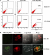
FIGURE 1.
FIGURE 1.
Hsp70L1 binds 293-TLR4 on the cell surface. A, HEK 293 cells and 293-TLR4 cells were incubated with 2 μg/ml PE-TLR4 or 5 μg/ml FITC-labeled Hsp70L1 for 30 min at 4 °C. After washing, the cells were analyzed by flow cytometry. Column 1, isotype controls (HEK 293 cells, upper, and 293-TLR4 cells, lower); column 2, HEK 293 cells (upper) and 293-TLR4 cells (lower) stained with PE-conjugated anti-TLR4 mAb; column 3, HEK 293 cells(upper) and 293-TLR4 cells (lower) incubated with FITC-labeled Hsp70L1. B, HEK 293 cells and 293-TLR4 cells were harvested and cultured on coverslips at the density of 50%, and then transiently transfected with Mem-DsRed vector. After 24 h, the cells were fixed by 4% paraformaldehyde for 30 min at 4 °C. After washing, the cells were stained with 2 μg/ml PE-TLR4 or 5 μg/ml FITC-labeled Hsp70L1 and analyzed by confocal microscope. Green fluorescence represents FITC-labeled Hsp70L1, and red fluorescence represents the cell membrane.
FIGURE 2.
FIGURE 2.
Activation of signaling pathways in 293-TLR4 cells by Hsp70L1. HEK293 cells and 293-TLR4 cells were stimulated with 10 μg/ml Hsp70L1 for the indicated time. Phospho-ERK, phospho-38, phospho-JNK, phospho-IKKα/β, and phosphor-IκB-α as well as total ERK, p38, JNK, IKKα, and IκB-α were detected by immunoblot.
FIGURE 3.
FIGURE 3.
Binding of Hsp70L1 to membrane TLR4 of DCs. A, GST-Hsp70L1 fusion protein was purified and assessed by SDS-PAGE. B, for lane 1 to lane 3, GST-Hsp70L1 fusion proteins and GST protein were combined to glutathione-beads, and incubated with membrane protein (1.1 mg/ml, 200 μl) prepared from immature DC. After washing with 20 μl of buffer, complexes were released by boiling, electrophoresed on 10% SDS-PAGE, and subjected to immunoblot with anti-TLR4 antibody. For lane 4, membrane protein complexes (1.1 mg/ml, 20 μl) prepared from immature DC were released by boiling, electrophoresed on 10% SDS-PAGE, and subjected to immunoblot with anti-TLR4 antibody to identify TLR4 existence.
FIGURE 4.
FIGURE 4.
TLR4 is required for phenotypic and functional maturation of mouse DCs induced by Hsp70L1. A, 5 day bone marrow-derived DCs (5 × 10⁵/ml) were stimulated with 10 μg/ml Hsp70L1 for 48 h, then collected for FACS analysis of CD40, CD80, CD86, and I-ab expression. Gray histograms indicate negative control; open thin line histograms, unstimulated DCs; and open thick line histograms, Hsp70L1-stimulated DCs. Experiments were performed independently at least three times. B–D, cytokine production of DCs stimulated with Hsp70L1 protein. Wild type DCs (WT) and TLR4-deficient DCs (TLR4−/−) were cultured for 5 days and stimulated with or without 10 μg/ml Hsp70L1 respectively for 6, 24, and 48 h. The levels of TNF-α, IL-1β, and IL-12p70 in the supernatants were measured by ELISA. Results are presented as mean S.D. of triplicate samples. (ND, not detected) (**, p < 0.01).
FIGURE 5.
FIGURE 5.
TLR4 mediates in vivo induction of specific Th1 immune responses by immunization with Hsp70L1-CEA576–669-pulsed DCs. Wild type (WT) mice and TLR4-deficient (TLR4−/−) mice were immunized with syngeneic DCs pulsed with Hsp70L1-CEA576–669, Hsp70L1, or CEA576–669. Then splenocytes were isolated and used to assay specific Th1 immune responses by IFN-γ ELISPOT (A) and Granzyme B ELISPOT (B). The results were indicated by the number positive SFCs/2 × 105 splenocytes. Columns, mean of three independent experiments; bars, S.E. **, p < 0.01.
FIGURE 6.
FIGURE 6.
Human DCs pulsed with Hsp70L1-CEA576–669 induces specific T cell response through TLR4-dependent manner. Human PBLs from HLA-A2.1+ healthy donors were stimulated with autologous DCs pulsed with Hsp70L1-CEA576–669, Hsp70L1, or CEA576–669. For TLR4 blockade experiment, the autologous DCs were blocked with 30 μg/ml TLR4 antagonist HAT125 1 h before DC pulsing. The IFN-γ (A) and Perforin ELISPOT (B) were used to analyze Th1 immune response. The results were indicated by the number positive SFCs/2 × 105 lymphocytes. Columns, mean of three independent experiments; bars, S.E. **, p < 0.01.

References

    1. Takeuchi O., Akira S. (2010) Cell 140, 805–820 - PubMed
    1. Kawai T., Akira S. (2010) Nat. Immunol. 11, 373–384 - PubMed
    1. Murata M. (2008) Cancer Sci. 99, 1435–1440 - PMC - PubMed
    1. Lundberg A. M., Drexler S. K., Monaco C., Williams L. M., Sacre S. M., Feldmann M., Foxwell B. M. (2007) Blood 110, 3245–3252 - PubMed
    1. Peter M. E., Kubarenko A. V., Weber A. N., Dalpke A. H. (2009) J. Immunol. 182, 7690–7697 - PubMed

Publication types