. 2011 Sep 2;286(35):30455-30461.
doi: 10.1074/jbc.M111.247338. Epub 2011 Jul 5.

Peroxisomes are involved in biotin biosynthesis in Aspergillus and Arabidopsis

Affiliations

Peroxisomes are involved in biotin biosynthesis in Aspergillus and Arabidopsis

Yasuko Tanabe et al. J Biol Chem. .

Abstract

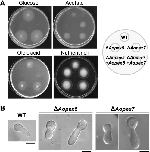
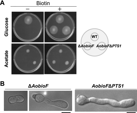
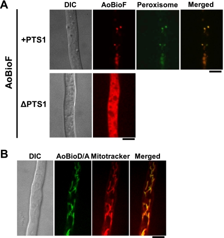
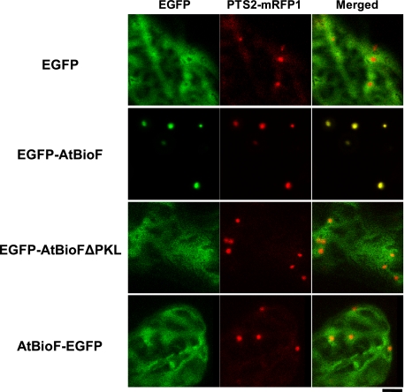
Among the eukaryotes only plants and a number of fungi are able to synthesize biotin. Although initial events leading to the biosynthesis of biotin remain largely unknown, the final steps are known to occur in the mitochondria. Here we deleted the Aopex5 and Aopex7 genes encoding the receptors for peroxisomal targeting signals PTS1 and PTS2, respectively, in the filamentous fungus Aspergillus oryzae. In addition to exhibiting defects in the peroxisomal targeting of either PTS1 or PTS2 proteins, the deletion strains also displayed growth defects on minimal medium containing oleic acid as the sole carbon source. Unexpectedly, these peroxisomal transport-deficient strains also exhibited growth defects on minimal medium containing glucose as the sole carbon source that were remediated by the addition of biotin and its precursors, including 7-keto-8-aminopelargonic acid (KAPA). Genome database searches in fungi and plants revealed that BioF protein/KAPA synthase, one of the biotin biosynthetic enzymes, has a PTS1 sequence at the C terminus. Fungal ΔbioF strains expressing the fungal and plant BioF proteins lacking PTS1 still exhibited growth defects in the absence of biotin, indicating that peroxisomal targeting of KAPA synthase is crucial for the biotin biosynthesis. Furthermore, in the plant Arabidopsis thaliana, AtBioF localized to the peroxisomes through recognition of its PTS1 sequence, suggesting involvement of peroxisomes in biotin biosynthesis in plants. Taken together we demonstrate a novel role for peroxisomes in biotin biosynthesis and suggest the presence of as yet unidentified peroxisomal proteins that function in the earlier steps of biotin biosynthesis.


Figures

FIGURE 1.
FIGURE 1.
Growth impairment of strains defective in peroxisomal targeting signal receptors on glucose minimal medium. A, growth of the A. oryzae ΔAopex5 and ΔAopex7 strains on minimal media (CD + Met) containing either glucose or acetate or oleic acid as the sole carbon source, and nutrient-rich medium (DPY). Conidial solutions (103 conidia/5 μl) of the wild-type, ΔAopex5, and ΔAopex7 strains and the complemented strains were spotted on each medium (as indicated to the right) and incubated at 30 °C for 3 days. B, swollen morphology of germinated conidia of the ΔAopex5 and ΔAopex7 strains in glucose minimal medium. Conidia of the wild-type, ΔAopex5, and ΔAopex7 strains were inoculated in minimal medium (CD + Met) containing glucose at a concentration of 103 conidia/100 μl. The cultures were incubated at 30 °C for 7 h (wild-type) or 15 h (ΔAopex5 and ΔAopex7) and observed by microscopy. Note that a longer incubation time was required for the ΔAopex5 and ΔAopex7 strains to germinate. Bars: 5 μm.
FIGURE 2.
FIGURE 2.
Biotin and its biosynthetic precursors restore the growth defects of the peroxisome-deficient strains on glucose minimal medium. Conidial solutions (103 conidia/5 μl) of the wild-type, ΔAopex5, and ΔAopex7 strains were spotted on minimal medium (CD + Met) containing glucose supplemented with biotin or its biosynthetic precursors, pimelic acid (Pim), pimeloyl-CoA (Pim-CoA), 7-keto-8-aminopelargonic acid (KAPA), and dethiobiotin (DTB). Photographs of the cultures were taken after incubation at 30 °C for 3 days.
FIGURE 3.
FIGURE 3.
Deletion of PTS1 from BioF protein leads to biotin auxotrophy on glucose minimal medium. A, growth of the ΔAobioF and AobioFΔPTS1 strains on minimal medium (M + Met) containing either glucose or acetate as the sole carbon source supplemented with biotin. Conidia solutions (103 conidia/5 μl) of the strains were spotted on plates (as indicated to the right) and incubated at 30 °C for 3 days. B, swollen morphology of germinated conidia of the ΔAobioF and AobioFΔPTS1 strains in glucose minimal medium. Conidia of these strains were inoculated in minimal medium (CD + Met) containing glucose and 0.5% casamino acids at a concentration of 103 conidia/100 μl. Photographs of germinated conidia were taken after the cultures were incubated at 30 °C for 20 h. Bars: 5 μm.
FIGURE 4.
FIGURE 4.
Localization analysis of AoBioF and its downstream enzyme. A, AoBioF localization in peroxisomes. Conidia of strains expressing either AoBioF-mDsRed-PTS1 or AoBioF-mDsRed-ΔPTS1 were inoculated in minimal medium (CD + Met) containing glucose and 0.5% casamino acids. After a 20-h incubation at 30 °C, the hyphae were observed by fluorescence microscopy. To visualize peroxisomes, EGFP-PTS1 was also expressed in the A. oryzae strain expressing AoBioF-mDsRed-PTS1. Bars: 5 μm. B, AoBioD/A localization in mitochondria. The strain expressing AoBioD/A-EGFP endogenously was grown at 30 °C for 24 h in minimal medium (CD + Met) containing glucose and 0.5% casamino acids. Mitochondria were stained with MitoTracker Red CMXRos and observed by fluorescence microscopy. DIC, differential interference contrast microscopy. Bar: 5 μm.
FIGURE 5.
FIGURE 5.
Plant BioF localizes to peroxisomes by utilizing the PTS1 sequence at the C terminus. Confocal images of EGFP (EGFP) and PTS2-mRFP1 fluorescence (PTS2-mRFP1) and their merged images (Merged) are shown in Arabidopsis leaf epidermal cells transiently expressing the cDNA constructs of EGFP (EGFP) and the full-length (EGFP-AtBioF) and C-terminal proline-lysine-leucine-deleted mutant proteins of AtBioF (EGFP-AtBioFΔPKL) fused to the C terminus of EGFP. The full-length AtBioF fused to the N terminus of EGFP (AtBioF-EGFP) was also examined. Bar: 5 μm.
FIGURE 6.
FIGURE 6.
Schematic model of eukaryotic biotin biosynthesis. BioF is transported into peroxisomes through the recognition of the PTS1 motif by Pex5, and it converts pimeloyl-CoA to KAPA. Our study suggests that a PTS2-dependent protein participates in the supply of pimelic acid and a PTS1-dependent protein then synthesizes pimeloyl-CoA from pimelic acid. Note that in acetate minimal medium BioFΔPTS1 is not transported to peroxisomes; it synthesizes KAPA from pimeloyl-CoA supplied in the cytoplasm (see “Discussion”).

References

    1. Wanders R. J., Waterham H. R. (2006) Annu. Rev. Biochem. 75, 295–332 - PubMed
    1. Mano S., Nishimura M. (2005) Vitam. Horm. 72, 111–154 - PubMed
    1. Reumann S., Weber A. P. (2006) Biochim. Biophys. Acta 1763, 1496–1510 - PubMed
    1. Weber H. (2002) Trends Plant Sci. 7, 217–224 - PubMed
    1. Woodward A. W., Bartel B. (2005) Ann. Bot. 95, 707–735 - PMC - PubMed

Publication types