. 2011 Jul 19;108(29):11924-9.
doi: 10.1073/pnas.1103418108. Epub 2011 Jul 5.

Ezrin-mediated apical integrity is required for intestinal homeostasis

Affiliations

Ezrin-mediated apical integrity is required for intestinal homeostasis

Jessica B Casaletto et al. Proc Natl Acad Sci U S A. .

Abstract

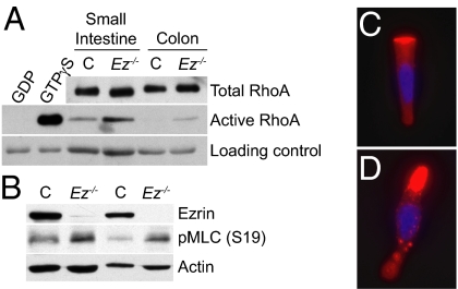
Individual cell types are defined by architecturally and functionally specialized cortical domains. The Ezrin, Radixin, and Moesin (ERM) proteins play a major role in organizing cortical domains by assembling membrane protein complexes and linking them to the cortical actin cytoskeleton. Many studies have focused on the individual roles of the ERM proteins in stabilizing the membrane-cytoskeleton interface, controlling the distribution and function of apical membrane complexes, regulating the small GTPase Rho, or establishing cell-cell junctions. We previously found that deletion of the mouse Ezrin gene yields severe defects in apical integrity throughout the developing intestinal epithelium, resulting in incomplete villus morphogenesis and neonatal death. However, the molecular function of Ezrin in building the apical surface of the intestinal epithelium was not clear. By deleting Ezrin in the adult mouse intestinal epithelium, we provide evidence that Ezrin performs multiple molecular functions that collaborate to build the functional apical surface of the intestinal epithelium in vivo. The loss of Ezrin-mediated apical integrity in the adult intestine yields severe morphological consequences during intestinal homeostasis, including defects in cell geometry, extrusion, junctional remodeling, and spindle orientation. Surprisingly, deletion of Ezrin either before or after villus morphogenesis yields villus fusion, revealing a previously unrecognized step in intestinal homeostasis. Our studies indicate that the function of Ezrin in building and maintaining the apical domain is essential not only for intestinal morphogenesis but also for homeostasis in the mature intestine.


Conflict of interest statement

The authors declare no conflict of interest.

Figures

Fig. 1.
Fig. 1.
Apical defects in the adult Ez−/− intestine. (AD) Transmission EM of intestinal epithelia from tamoxifen-treated Ezlox/lox (A and C) and Vil-Cre-ERT2;Ezlox/lox (B and D) mice. Control colonic (A) and small intestinal (C) epithelia maintain a highly organized BB featuring densely packed, uniform microvilli with actin rootlets embedded in a compact terminal web. Ez−/− microvilli in colonic (B) and small intestinal epithelia (D) are nonuniform, misoriented, and extend from a thickened and disorganized terminal web; actin rootlets are disorganized or absent. (A and B) 2,900× magnification. (C and D) 11,000× magnification. (E and F) Large blebs of membrane no longer attached to the cytoskeleton are frequently found throughout the Ez−/− intestine. (E) 15,000× magnification. (F) 7,100× magnification. (G and H) Phalloidin-stained colonic epithelia reveal the continuous apical band of actin labeling of the BB of control cells (G); actin is discontinuous in Ez−/− cells (H). (G and H) 600× magnification.
Fig. 2.
Fig. 2.
Loss of Ezrin disrupts the formation of apical membrane complexes. (A) BBs were isolated from control (lanes 1 and 3) and Ez−/− (lanes 2 and 4) small intestines. Total cell lysate (TCL) and BB fractions were solubilized in the presence of 1% SDS. Villin served as a positive control for the BB fraction and aminopeptidase N (APN), and Crumbs3 served as controls for apical membrane proteins. Note the specific loss of NHERF1 and Slk in Ez−/− BBs. (B and C) NHERF1 is apically concentrated in colonic epithelia from tamoxifen-treated Ezlox/lox (control, B), but not Vil-Cre-ERT2;Ezlox/lox (Ez−/−, C) mice.
Fig. 3.
Fig. 3.
Increased Rho activity in Ez−/− intestinal epithelial cells. (AD) Cells from vehicle (C) or tamoxifen-treated (Ez−/−) Vil-Cre-ERT2;Ezlox/lox mice were analyzed. (A) Cell lysates were incubated with GST-Rhotekin beads. Bound proteins (active) and total cell lysates (total) were examined. Preincubation with GDP or GTPγS served as negative and positive controls, respectively. The amount of beads in each reaction served as a loading control for each pair. (B) Small intestinal epithelial cell lysates from two pairs of C and Ez−/− mice reveal increased levels of pMLC in Ez−/− cells. (C and D) Control (C) or Ez−/− (D) small intestinal epithelial cells were immunostained with antibodies against pMLC (Ser19) (red) and DAPI (blue). In Ez−/− cells, pMLC is enriched in the thickened apical region and present in punctate aggregates (D). (C and D) 600× magnification.
Fig. 4.
Fig. 4.
Morphogenetic consequences of Ezrin loss in the adult intestine. (AC) Altered shape of Ez−/− epithelial cells. (A and B) β-Catenin staining in colonic epithelia from tamoxifen-treated Ezlox/lox (control, A) and Vil-Cre-ERT2;Ezlox/lox (Ez−/−, B) mice reveals expanded apical surfaces of control cells as they transition from the crypt to the colonic surface (A) in contrast to the nearly uniformly narrow Ez−/− cells (B). An asterisk marks the apical surfaces that compose this transition and provides an example of the quantitation shown in C. (A and B) 600× magnification. (C) The number of apical surfaces per transition in control (C) (4.0 ± 0.6) and Ez−/− (7.6 ± 1.3) mice were determined using two mice of each genotype (P < 0.0001). (DF) Decreased extrusion of apoptotic cells in Ez−/− epithelia. Cleaved caspase-3 staining of control (E) and Ez−/− (F) epithelia reveals a sixfold increase in positive cells at the colonic surface in the absence of Ezrin (control, 0.64% ± 0.25; Ez−/−, 3.8% ± 0.28; P < 0.01). A minimum of 1,000 epithelial cells per colon were counted using two mice of each genotype. (G and H) SEM across the luminal surface of the colon. Apical defects result in a rough, nonuniform appearance of the Ez−/− colon (H) compared with the smooth, uniform surface of the control (G). (G and H) 130× magnification. (I and J) SEM across the luminal surface of the small intestinal epithelium from control (I) and Ez−/− (J) mice. Single-villus units cover the control small intestine, whereas Ez−/− intestines contain villus structures composed of two or more individual villi fused together. (I and J) 250× magnification.
Fig. 5.
Fig. 5.
Defects in the AJR of Ez−/− intestinal epithelia. (A and B) Transmission EM of colonic epithelia from tamoxifen-treated Ezlox/lox (control, A) and Vil-Cre-ERT2;Ezlox/lox (Ez−/−, B) mice reveals defective AJR architecture in the absence of Ezrin. (A and B) 11,000× magnification. (C) Increased solubility of AJ components in the Ez−/− BB. AJ components in the total cell lysate (TCL) and BB fractions of control (lanes 1, 3, 5, and 7) and Ez−/− (lanes 2, 4, 6, and 8) small intestinal epithelia were solubilized in the presence of 1% or 0.1% SDS. Note the increased solubility in Ez−/− BBs. The solubilities of the tight junction proteins ZO-1 and occludin were also increased (Fig. S7). (D) Co-immunoprecipitation of β-catenin and α-catenin with E-cadherin in the indicated sucrose gradient fractions (Fig. S6) reveals the presence of higher-molecular-weight AJ complexes in Ez−/− intestines.
Fig. 6.
Fig. 6.
Altered spindle orientation in the absence of Ezrin. (A and B) Immunostaining with anti-pericentrin antibodies (green), phalloidin (red, marks the crypt–villus axis), and DAPI (blue) reveals spindle orientation in small intestinal crypts from tamoxifen-treated Ezlox/lox (control, A) and Vil-Cre-ERT2;Ezlox/lox (Ez−/−, B) mice. Representative single z-plane images are presented. Note that both centrosomes are found in a single z-plane in control crypts (A), whereas the two centrosomes in B are in distinct z-planes that are 4 μm apart. (C) Quantitation of spindle orientation carried out by confocal imaging and 3D reconstruction reveals that 91% ± 7.5% (n = 23; P < 0.0005) of spindles in control crypts were aligned within 30° of the crypt–villus axis, whereas only 67% ± 8.0% (n = 30; P < 0.05) of spindles in Ez−/− crypts were aligned in this manner (overall P < 0.05).

References

    1. McClatchey AI, Fehon RG. Merlin and the ERM proteins: Regulators of receptor distribution and signaling at the cell cortex. Trends Cell Biol. 2009;19:198–206. - PMC - PubMed
    1. Fehon RG, McClatchey AI, Bretscher A. Organizing the cell cortex: The role of ERM proteins. Nat Rev Mol Cell Biol. 2010;11:276–287. - PMC - PubMed
    1. Algrain M, Turunen O, Vaheri A, Louvard D, Arpin M. Ezrin contains cytoskeleton and membrane binding domains accounting for its proposed role as a membrane-cytoskeletal linker. J Cell Biol. 1993;120:129–139. - PMC - PubMed
    1. Pearson MA, Reczek D, Bretscher A, Karplus PA. Structure of the ERM protein moesin reveals the FERM domain fold masked by an extended actin binding tail domain. Cell. 2000;101:259–270. - PubMed
    1. Reczek D, Berryman M, Bretscher A. Identification of EBP50: A PDZ-containing phosphoprotein that associates with members of the ezrin-radixin-moesin family. J Cell Biol. 1997;139:169–179. - PMC - PubMed

Publication types