

Apolipoprotein A1 and C-terminal fragment of α-1 antichymotrypsin are candidate plasma biomarkers associated with acute renal allograft rejection
- PMID: 21730889
- PMCID: PMC3878300
- DOI: 10.1097/TP.0b013e318225db6a
Apolipoprotein A1 and C-terminal fragment of α-1 antichymotrypsin are candidate plasma biomarkers associated with acute renal allograft rejection
Abstract
Background: Current diagnostic methods of renal allograft rejection are neither sensitive nor specific. Needle biopsies are invasive and associated with patient morbidity. Thus, it is desirable to develop noninvasive tests to predict and diagnose rejection.
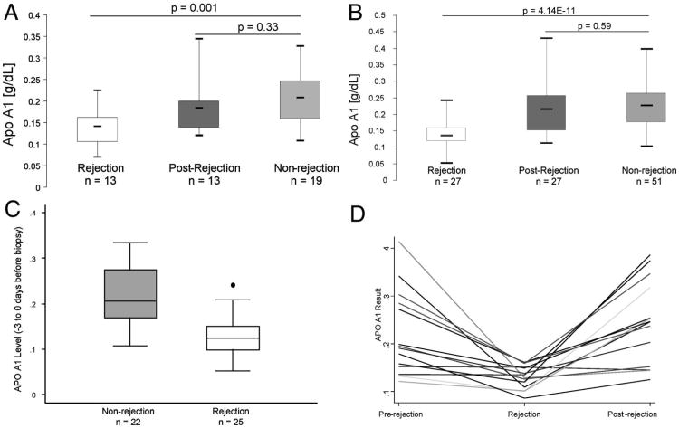
Methods: Using a case-control approach, surface-enhanced laser desorption/ionization time-of-flight mass spectrometry was used to identify plasma proteins associated with renal allograft rejection. From each rejection patient (n=16), two plasma samples (one near the biopsy date and the other at a time postbiopsy) were compared. Biopsy-confirmed nonrejection patients (n=48) were further analyzed as controls. Antibody-based quantitative enzyme-linked immunosorbent assay was performed to validate candidate biomarker apolipoprotein A1 (Apo A1) in a subset of the original and a second cohort of biopsy-confirmed rejection (n=40) and nonrejection (n=70) patients.
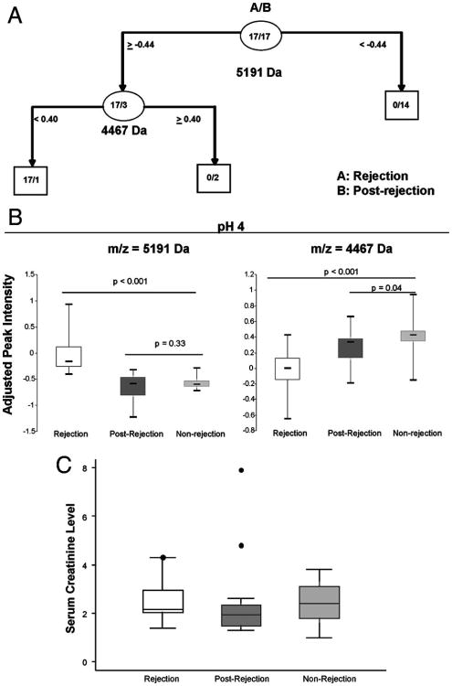
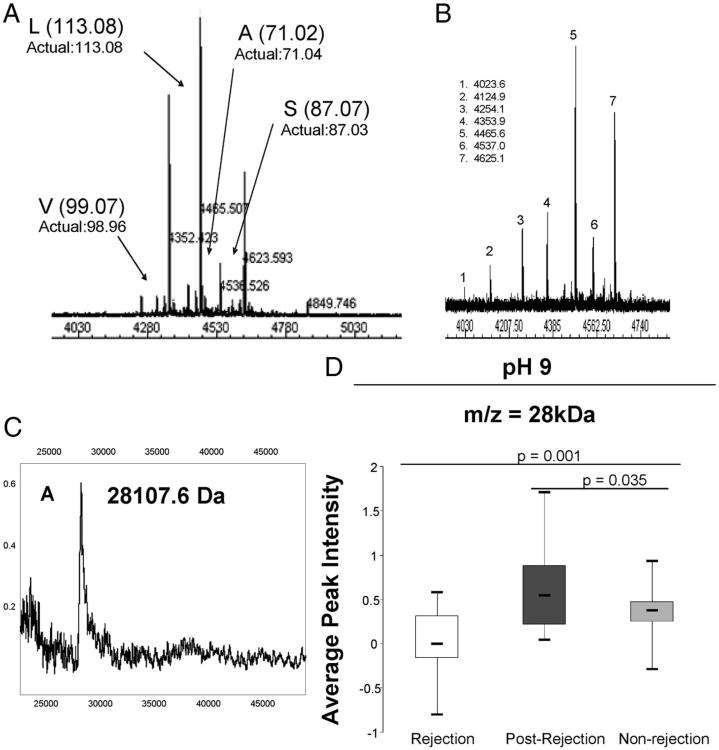
Results: Twenty-two proteins/peptides showed significant differences between rejection and postrejection samples. Peptides 5191 Da and 4467 Da detected rejection with 100% sensitivity and 94% specificity. The 4467 Da peptide was identified as the C-terminal fragment of α-1 antichymotrypsin and a 28 kDa protein was determined as Apo A1. Both protein levels were significantly lower at rejection compared with postrejection. Protein levels of nonrejection patients were similar to the postrejection samples. Apo A1 enzyme-linked immunosorbent assay results showed significantly lower Apo A1 levels (P=0.001 for the original and P=4.14E-11 for the second cohort) at the time of rejection compared with nonrejection which coincides with the SELDI findings.
Conclusions: Together α-1 antichymotrypsin, Apo A1, and the unidentified 5191 Da peptide provide a plasma molecular profile, and this is associated with acute cellular renal allograft rejection.
Conflict of interest statement
The authors declare no conflicts of interest.
Figures
References
-
- Li B, Hartono C, Ding R, et al. Noninvasive diagnosis of renal-allograft rejection by measurement of messenger RNA for perforin and granzyme B in urine. N Engl J Med. 2001;344:947. - PubMed
-
- Clarke W. Proteomic research in renal transplantation. Ther Drug Monit. 2006;28:19. - PubMed
-
- Beckingham IJ, Nicholson ML, Bell PR. Analysis of factors associated with complications following renal transplant needle core biopsy. Br J Urol. 1994;73:13. - PubMed
-
- Rush DN, Henry SF, Jeffery JR, et al. Histological findings in early routine biopsies of stable renal allograft recipients. Transplantation. 1994;57:208. - PubMed
Publication types
MeSH terms
Substances
Grants and funding
LinkOut - more resources
Full Text Sources
Other Literature Sources
Medical
