. 2011 Sep;31(9):e1-10.
doi: 10.1038/jcbfm.2011.96. Epub 2011 Jul 6.

Metabotropic glutamate receptor mGluR5 is not involved in the early hemodynamic response

Affiliations

Metabotropic glutamate receptor mGluR5 is not involved in the early hemodynamic response

Novella Calcinaghi et al. J Cereb Blood Flow Metab. 2011 Sep.

Abstract

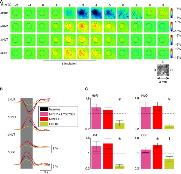
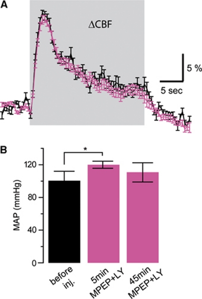
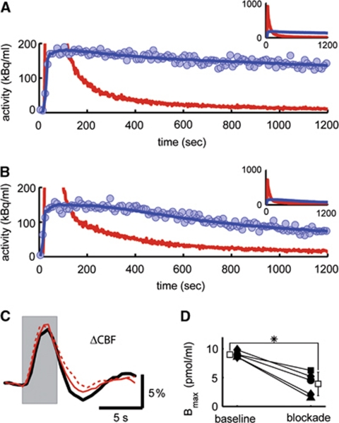
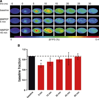
Activation of astrocytic metabotropic glutamate receptor 5 (mGluR5) is postulated to elicit calcium transients, triggering a chain of events that ultimately regulates cerebral blood flow by changing the tone of smooth muscle cells of nearby arterioles. Using concurrent in vivo optical imaging and determination of receptor occupancy with (11)C-ABP688, we report here that blocking ∼80% of mGluR5 in vivo does not affect transient hemodynamic responses on brief whisker stimulations while transiently reducing neuronal activity as measured by voltage-sensitive dye imaging. Our results show that mechanisms other than activation of mGluR5 are required to trigger the initial hemodynamic response in normal physiological conditions.


Figures

Figure 1
Figure 1
Application of group I metabotropic glutamate receptor (mGluR) antagonists does not affect neurovascular coupling on brief whisker stimulation. (A) Relative changes of deoxy- (HbR), oxyhemoglobin (HbO), and blood volume (HbT) on brief whisker stimulation were measured by optical spectroscopy in the activated barrel cortex, whereas cerebral blood flow (CBF) was measured by laser speckle imaging. (B) Average responses after intravenous injection of the mGluR5 and mGluR1 antagonists 6-methyl-2-(phenylethynyl)-pyridine (MPEP) and LY367385, respectively (1.2 mg/kg; pink), and after injection of 2-(3-methoxy-phenylethynyl)-6-methyl-pyridine (M-MPEP; four animals at 1 mg/kg; one animal at 2 mg/kg; one animal at 3 mg/kg; and two animals at 4 mg/kg; red) compared with baseline (black). Data shown were recorded 10 minutes after drug injection. No significant decrease of the response was observed after these treatments (statistical testing either for the MPEP–LY367385 and M-MPEP groups separately or for the two groups pooled together, n=15). In contrast, after treatment with 6-cyano-7-nitroquinoxaline-2,3-dione (CNQX; 15 mg/kg; green; n=6), a strong reduction in the hemodynamic signals was evident. (C) Peak deviations normalized to baseline (mean±s.e.m.; *P<0.05, P=0.075, Wilcoxon signed-rank test). In total, MPEP–LY367385, M-MPEP, and CNQX were tested, respectively, in 7, 8, and 6 rats. Measurements at the later time points (30 and 45 minutes) did not show any significant decrease of the peak response (data not shown).
Figure 2
Figure 2
The neurovascular coupling response during longer stimulation is also not affected by group I metabotropic glutamate receptor antagonists 6-methyl-2-(phenylethynyl)-pyridine (MPEP) and LY367385. (A) Example of cerebral blood flow (CBF) recordings during 24-second whisker stimulation before (black) and 10 minutes after (pink) MPEP and LY367385 intravenous injection (4.0 mg/kg). (B) Systemic effect on mean arterial blood pressure (MAP) evoked by MPEP and LY367385 intravenous injection (4.0 mg/kg). Five minutes after drug injection, a significant increase was observed (MAP before injection: 101±11 mm Hg; 5 minutes after injection: 120±4 mm Hg; 45 minutes after injection: 111±11 mm Hg; mean±s.d.; n=4; *P<0.05, Wilcoxon signed-rank test) before the arterial blood pressure returned to its baseline. This transient increase was not elicited by placebo injection (see text).
Figure 3
Figure 3
Delivery of 2-(3-methoxy-phenylethynyl)-6-methyl-pyridine (M-MPEP) leads to blockage of metabotropic glutamate receptor 5 (mGluR5) in the rat somatosensory cortex but fails to attenuate the hemodynamic response. After the injection of M-MPEP (4 mg/kg), the washout of radiolabel from tissue was increased (B) compared with baseline condition (A) as represented by the decay-corrected tissue time–activity curves (blue circles). The model curve (blue line) and the radioactivity concentration in plasma (that is input curve; red line) are also shown. In A and B, the ordinate is truncated at 200 kBq/mL for legibility reasons. The full range of data is displayed in the insets (same axes legends). In this example, the estimated mGluR5 blockage amounted to 47%. (C) Hemodynamic response on stimulation (gray area) before (black line), 10 minutes (red line), and 30 minutes (broken red line) after delivery of M-MPEP (same experiment as in A and B) was unaffected by mGluR5 blockage. (D) The density of receptors in the tissue (Bmax) is shown for all individual β-probe experiments before and after blockage (square: 1.2 mg/kg 6-methyl-2-(phenylethynyl)-pyridine/LY367385; circle: 1 mg/kg M-MPEP; diamond: 4 mg/kg M-MPEP; and triangle: 8 mg/kg M-MPEP). White boxes represent mean±s.d. (*P<0.05, Wilcoxon signed-rank test).
Figure 4
Figure 4
Neuronal activity slightly decreases during the first 30 minutes after metabotropic glutamate receptor 5 blockage. (A) Maps of a single voltage-sensitive dye (VSD) imaging experiment performed before (top), 5 minutes (middle), and 45 minutes (bottom) after injection of 2-(3-methoxy-phenylethynyl)-6-methyl-pyridine (M-MPEP; 4 mg/kg). Single frames are 5 milliseconds apart from each other. The series starts 5 milliseconds before single whisker stimulation evoked at time 0. (B) Summary of VSD results in six animals. After only 5 minutes after the injection of M-MPEP (4 mg/kg), the amplitude of the VSD signal significantly decreased by, on average, 27%. At later time points, the signal was not significantly different from baseline level; *P<0.05, Wilcoxon signed-rank test.

References

    1. Aronica E, van Vliet EA, Mayboroda OA, Troost D, da Silva FH, Gorter JA. Upregulation of metabotropic glutamate receptor subtype mGluR3 and mGluR5 in reactive astrocytes in a rat model of mesial temporal lobe epilepsy. Eur J Neurosci. 2000;12:2333–2344. - PubMed
    1. Attwell D, Buchan AM, Charpak S, Lauritzen M, MacVicar BA, Newman EA. Glial and neuronal control of brain blood flow. Nature. 2010;468:232–243. - PMC - PubMed
    1. Ballester-Rosado CJ, Albright MJ, Wu CS, Liao CC, Zhu J, Xu J, Lee LJ, Lu HC. mGluR5 in cortical excitatory neurons exerts both cell-autonomous and -nonautonomous influences on cortical somatosensory circuit formation. J Neurosci. 2010;30:16896–16909. - PMC - PubMed
    1. Berwick J, Johnston D, Jones M, Martindale J, Martin C, Kennerley AJ, Redgrave P, Mayhew JEW. Fine detail of neurovascular coupling revealed by spatiotemporal analysis of the hemodynamic response to single whisker stimulation in rat barrel cortex. J Neurophysiol. 2008;99:787–798. - PMC - PubMed
    1. Biber K, Laurie DJ, Berthele A, Sommer B, Tolle TR, Gebicke-Harter PJ, van Calker D, Boddeke HWGM. Expression and signaling of group I metabotropic glutamate receptors in astrocytes and microglia. J Neurochem. 1999;72:1671–1680. - PubMed

Publication types

Substances

LinkOut - more resources