Chronic intestinal pseudo-obstruction in a horse: a case of myenteric ganglionitis
- PMID: 21731098
- PMCID: PMC3058658
Chronic intestinal pseudo-obstruction in a horse: a case of myenteric ganglionitis
Abstract
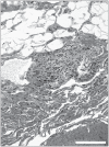
An 11-year-old Quarter horse mare was presented for recurrent episodes of colic. A chronic intestinal pseudo-obstruction was diagnosed. Medical treatment and surgical resection of the colon were performed but the condition did not improve and the horse was euthanized. Histopathological examination revealed a myenteric ganglionitis of the small intestine and ascending colon.
Pseudo-obstruction intestinale chronique chez une jument : un cas ganglionite myentérique. Une jument Quarter Horse âgée de 11 ans a été présentée pour coliques récurrentes. Une pseudo-obstruction intestinale chronique a été diagnostiquée. Le traitement médical et la résection chirurgicale du côlon n’ont amené aucune amélioration de la condition et l’animal a été euthanasié. L’examen histopathologique des tissus a révélé une ganglionite des plexus myentériques de l’intestin grêle et du colon ascendant.
(Traduit par les auteurs)
Figures


Similar articles
-
Equine myenteric ganglionitis: a case of chronic intestinal pseudo-obstruction.Cornell Vet. 1990 Jan;80(1):53-63. Cornell Vet. 1990. PMID: 2403425
-
Myenteric ganglionitis as a cause of recurrent colic in an adult horse.J Am Vet Med Assoc. 2012 Jun 15;240(12):1494-500. doi: 10.2460/javma.240.12.1494. J Am Vet Med Assoc. 2012. PMID: 22657934
-
Extensive myenteric ganglionitis in a case of equine chronic intestinal pseudo-obstruction associated with EHV-1 infection.J Comp Pathol. 2013 May;148(4):289-93. doi: 10.1016/j.jcpa.2012.07.004. Epub 2012 Aug 27. J Comp Pathol. 2013. PMID: 22935089
-
[Chronic idiopathic intestinal pseudo-obstruction: visceral myopathy. Report of 4 cases].Acta Gastroenterol Latinoam. 1993;23(4):239-43. Acta Gastroenterol Latinoam. 1993. PMID: 8203187 Review. Spanish.
-
[Subtotal colectomy in neoplastic obstruction of the left part of the colon].Wiad Lek. 1987 Mar 1;40(5):324-6. Wiad Lek. 1987. PMID: 3303699 Review. Polish. No abstract available.
Cited by
-
Chronic intestinal pseudo-obstruction associated with enteric ganglionitis in a Persian cat.JFMS Open Rep. 2016 Jun 14;2(1):2055116916655173. doi: 10.1177/2055116916655173. eCollection 2016 Jan-Jun. JFMS Open Rep. 2016. PMID: 28491428 Free PMC article.
-
Case report: Successful treatment of intestinal leiomyositis in a dog using adjunctive intravenous immunoglobulin.Front Vet Sci. 2024 Jul 23;11:1373882. doi: 10.3389/fvets.2024.1373882. eCollection 2024. Front Vet Sci. 2024. PMID: 39109347 Free PMC article.
-
Intestinal Leiomyositis: A Cause of Chronic Intestinal Pseudo-Obstruction in 6 Dogs.J Vet Intern Med. 2016 Jan-Feb;30(1):132-40. doi: 10.1111/jvim.13652. Epub 2015 Nov 26. J Vet Intern Med. 2016. PMID: 26608226 Free PMC article.
References
-
- Brown CC, Baker DC, Barker IK. Alimentary system. In: Maxie GM, editor. Jubb, Kennedy & Palmer’s Pathology of Domestic Animals. 5th ed. Vol. 2. Toronto, Ontario: Elsevier; 2007. pp. 86–90.
-
- Fenoglio-Preiser CM, Noffsinger AE, Lantz PE, Stemmermann GN, Isaacson PG. Gastrointestinal Pathology. 3rd ed. Philadephia: Lippincott Williams & Wilkins; 2008. Motility disorders; pp. 543–591.
-
- Harvey AM, Hall EJ, Day MJ, Moore AH, Battersby IA, Tasker S. Chronic intestinal pseudo-obstruction in a cat caused by visceral myopathy. J Vet Intern Med. 2005;19:111–114. - PubMed
-
- Eastwood JM, McInnes EF, White RN, Elwood CM, Stock G. Caecal impaction and chronic intestinal pseudo-obstruction in a dog. J Vet Med Series A. 2005;52:43–44. - PubMed
-
- Arrick RH, Kleine LJ. Intestinal pseudoobstruction in a dog. J Am Vet Med Assoc. 1978;172:1201–1205. - PubMed
Publication types
MeSH terms
LinkOut - more resources
Full Text Sources
