

Keratoameloblastoma of the mandible
- PMID: 21731268
- PMCID: PMC3125065
- DOI: 10.4103/0973-029X.72507
Keratoameloblastoma of the mandible
Abstract
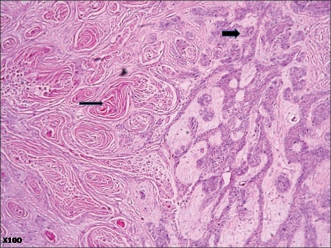
Keratoameloblastoma is a very rare ameloblastoma variant defined by extensive squamous metaplasia and keratinization. There are 13 previously reported cases in the literature, with a male predilection of 3:1. A 38-year-old male presented with a painless mandibular swelling which had been progressively increasing in size for 18 months. The incisional biopsy was misdiagnosed as basaloid squamous carcinoma. Owing to financial constraints, the patient had mandibular resection a decade after first noticing the growth, during which the clinical course was essentially benign, thus casting doubt on the initial diagnosis. The final histological diagnosis for both the incisional and resection biopsy specimens was keratoameloblastoma.
Keywords: Keratoameloblastoma; ameloblastoma; keratinization; mandible; misdiagnosis; prognosis.
Conflict of interest statement
Figures


References
-
- Gardner DG. Some current concepts on the pathology of ameloblastomas. Oral Surg Oral Med Oral Pathol Oral Radiol Endod. 1996;82:660–9. - PubMed
-
- Avon SL, Macomb J, Clokie C. Ameloblastic carcinoma: case report and literature review. J Can Dent Assoc. 2003;69:573–6. - PubMed
-
- Gilijamse M, Leemans CR, Winters HAH, Schulten EAJM, van der Waal I. Metastasizing ameloblastoma. Int J Oral Maxillofac Surg. 2007;36:462–4. - PubMed
-
- Van Rensburg LJ, Thompson IOC, Kruger HEC, Norval EJG. Hemangiomatous ameloblastoma: clinical, radiologic, and pathologic features. Oral Surg Oral Med Oral Pathol Oral Radiol Endod. 2001;9:374–80. - PubMed
