

Case Reports
doi: 10.4103/0973-029X.80014.
Aneurysmal bone cyst of the mandible: A case report and review of literature
Affiliations
- PMID: 21731290
- PMCID: PMC3125645
- DOI: 10.4103/0973-029X.80014
Item in Clipboard
Case Reports
Aneurysmal bone cyst of the mandible: A case report and review of literature
J Oral Maxillofac Pathol.
2011 Jan.
Abstract
Aneurysmal bone cyst (ABC) is rare benign lesions of bone which are infrequent in craniofacial skeleton. ABC's are characterized by rapid growth pattern with resultant bony expansion and facial asymmetry. We describe a case of ABC in a 25 year old male patient affecting the body of the mandible with expansion and thinning of the buccal and lingual cortical plates. Treatment consisted of surgical curettage of the lesion. A one year follow- up showed restoration of facial symmetry and complete healing of the involved site.
Keywords: Aneurysmal bone cyst; benign bone lesions; mandible; psuedocyst.
Conflict of interest statement
Figures
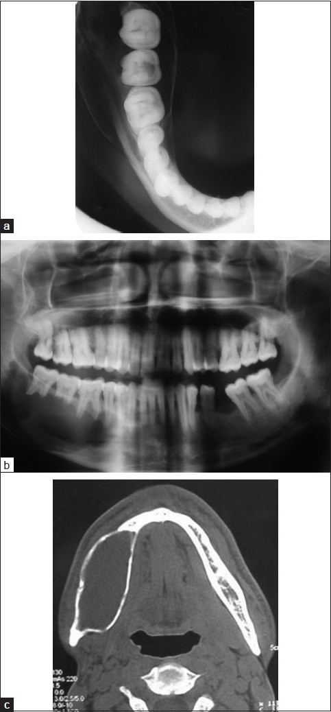
(a) Mandibular occlusal radiograph showing expansion of the cortical plates; (b) panoramic radiography showing a unilocular radiolucency extending from 45 to 48 region; (c) axial CT image of the mandible showing cortical expansion and thinning
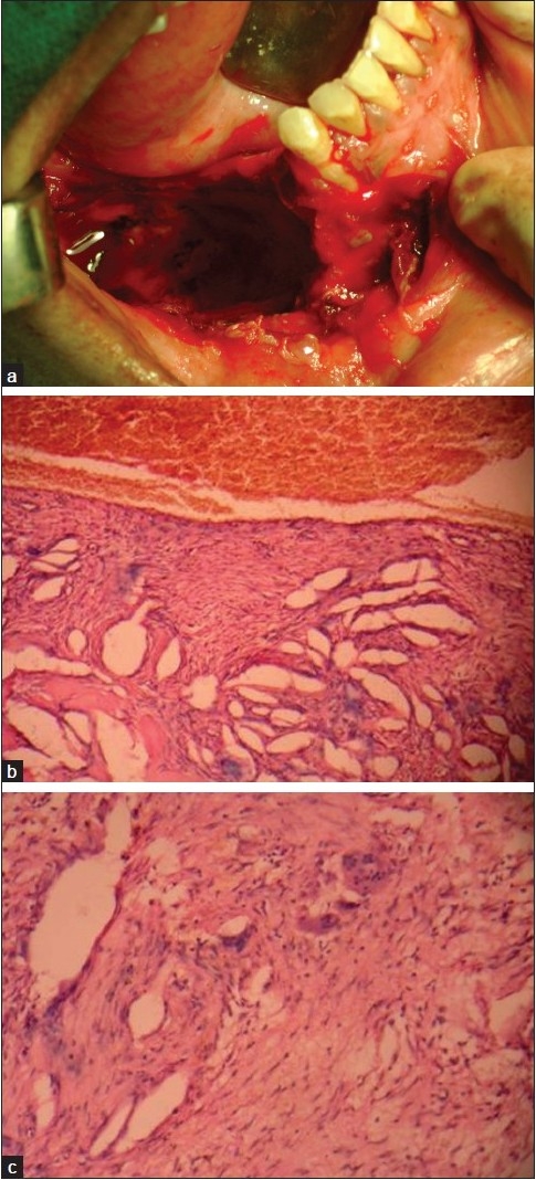
(a) Intraoperative photograph after the curettage; (b) photomicrograph of the lesion showing numerous vascular spaces lined by endothelial cells and multinucleated giant cells (H and E, 10×); (c) photomicrograph showing multinucleated giant cells in the fibrous tissue (H and E, 40×)
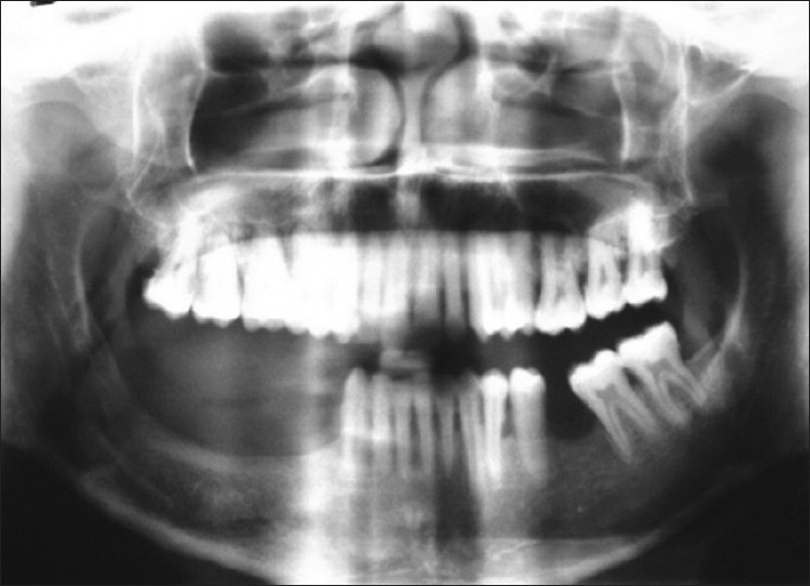
Postoperative panoramic radiograph, 1 year after curettage, showing no recurrence
Similar articles
-
Conversion of Aneurysmal Bone Cyst into Fibrous Dysplasia: A Rare Pediatric Case Report.J Maxillofac Oral Surg. 2016 Jul;15(Suppl 2):355-60. doi: 10.1007/s12663-016-0899-1. Epub 2016 Apr 20. J Maxillofac Oral Surg. 2016. PMID: 27408470 Free PMC article.
-
Dual Pathology of Mandible.Ann Maxillofac Surg. 2017 Jul-Dec;7(2):316-318. doi: 10.4103/ams.ams_146_16. Ann Maxillofac Surg. 2017. PMID: 29264307 Free PMC article.
-
Giant aneurysmal bone cyst of the mandible with unusual presentation.Med Oral Patol Oral Cir Bucal. 2009 Mar 1;14(3):E137-40. Med Oral Patol Oral Cir Bucal. 2009. PMID: 19242394
-
Mandibular aneurysmal bone cyst in a child misdiagnosed as acute osteomyelitis: a case report and a review of the literature.Eur J Pediatr. 2010 Aug;169(8):1037-40. doi: 10.1007/s00431-010-1138-2. Epub 2010 Jan 27. Eur J Pediatr. 2010. PMID: 20107833 Review.
-
Giant aneurysmal bone cyst of the mandible.J Craniofac Surg. 2011 Mar;22(2):745-8. doi: 10.1097/SCS.0b013e31820871b8. J Craniofac Surg. 2011. PMID: 21415654 Review.
Cited by
-
Imaging approach for jaw and maxillofacial bone tumors with updates from the 2022 World Health Organization classification.World J Radiol. 2024 Aug 28;16(8):294-316. doi: 10.4329/wjr.v16.i8.294. World J Radiol. 2024. PMID: 39239241 Free PMC article. Review.
-
A large aneurysmal bone cyst of mandible: A rare case report.J Oral Maxillofac Pathol. 2023 Jul-Sep;27(3):548-552. doi: 10.4103/jomfp.jomfp_459_22. Epub 2023 Sep 12. J Oral Maxillofac Pathol. 2023. PMID: 38033948 Free PMC article.
-
Odontogenic myxoma: A case report of a rare tumor.Radiol Case Rep. 2023 Sep 14;18(11):4130-4133. doi: 10.1016/j.radcr.2023.08.080. eCollection 2023 Nov. Radiol Case Rep. 2023. PMID: 37745767 Free PMC article.
-
Aneurysmal bone cyst of the mandible: a rare case report and literature review.Ann Med Surg (Lond). 2023 Aug 8;85(10):5133-5137. doi: 10.1097/MS9.0000000000001168. eCollection 2023 Oct. Ann Med Surg (Lond). 2023. PMID: 37811038 Free PMC article.
-
Role of FNAC in the diagnosis of intraosseous jaw lesions.Med Oral Patol Oral Cir Bucal. 2015 May 1;20(3):e284-91. doi: 10.4317/medoral.20274. Med Oral Patol Oral Cir Bucal. 2015. PMID: 25662547 Free PMC article.
References
-
- Gadre KS, Zubairy RA. Aneurysmal bone cyst of the mandibular condyle: Report of a case. J Oral Maxillofac Surg. 2000;58:439–43. - PubMed
-
- Motamedi MH, Stavropoulos M.F. Large radiolucent lesion of the mandibular condyle. J Oral Maxillofac Surg. 1997;55:1300–4. - PubMed
-
- Kalantar Motamedi MH. Aneurysmal bone cyst of the jaws: Clinicopathological features, radiographic evaluation and treatment analysis of 17 cases. J Craniomaxillofac Surg. 1998;26:56–62. - PubMed
-
- Rosenberg AE, Nielsen GP, Fletcher JA. Lyon: IARC Press; 2002. World Health Organisation Classification of tumours. Pathology and Genetics of Tumours of Soft tissues and Bone; p. 338.
