. 2011 Jun 1;303(2-3):191-198.
doi: 10.1016/j.ijms.2011.02.003.

Biologically Relevant Metal-Cation Binding Induces Conformational Changes in Heparin Oligosaccharides as Measured by Ion Mobility Mass Spectrometry

Affiliations

Biologically Relevant Metal-Cation Binding Induces Conformational Changes in Heparin Oligosaccharides as Measured by Ion Mobility Mass Spectrometry

Youjin Seo et al. Int J Mass Spectrom. .

Abstract

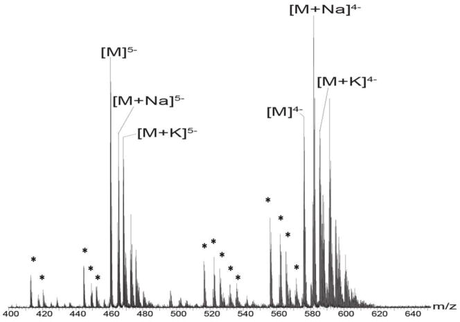
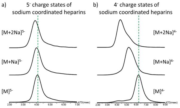
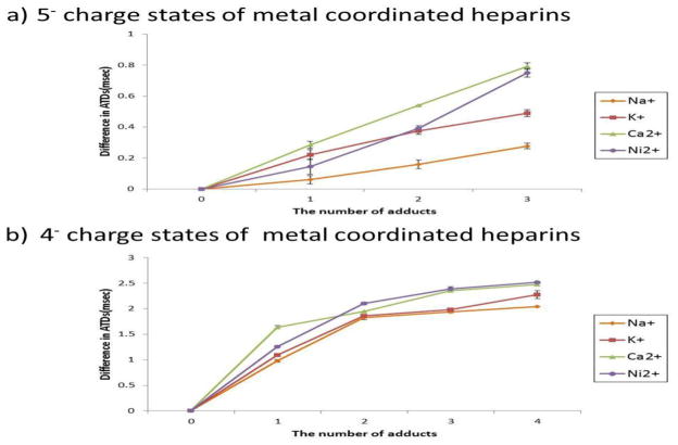
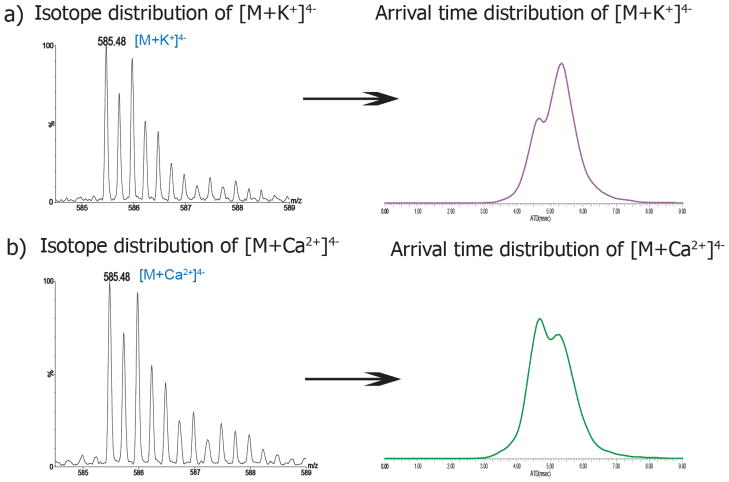
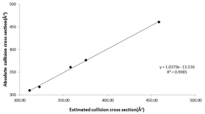
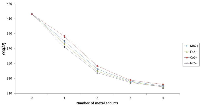
Heparin interacts with many proteins and is involved in biological processes such as anticoagulation, angiogenesis, and antitumorigenic activities. These heparin-protein interactions can be influenced by the binding of various metal ions to these complexes. In particular, physiologically relevant metal cations influence heparin-protein conformations through electronic interactions inherent to this polyanion. In this study, we employed ion mobility mass spectrometry (IMMS) to observe conformational changes that occur in fully-sulfated heparin octasaccharides after the successive addition of metal ions. Our results indicate that binding of positive counter ions causes a decrease in collision cross section (CCS) measurements, thus promoting a more compact octasaccharide structure.


Figures

Figure 1
Figure 1
Negative ion mode mass spectrum of 10μM dodecasulfated heparin octasaccharide dissolved in menthol/water/acetic acid solution (49/49/2; v/v/v). M represents dodecasulfated heparin octasaccharide. The charge distributions of 4- and 5- ions of heparin octasaccharides are illustrated. Asterisks represent desulfated ions corresponding to 5- or 4- charge state species. Mass to charge ratios of the heparin octasaccharides are shown in Table 1 of supplementary material.
Figure 2
Figure 2
The arrival time distributions for sodium coordinated heparin octasaccharide a) in the 5- charge state and b) in the 4- charge state. The dash lines indicate the baseline of the arrival time distribution for sodium-free coordinated heparin octasaccharide. Arrival time distributions are recorded in milliseconds.
Figure 3
Figure 3
The difference in arrival time distributions between metal and metal free coordinated heparin octasaccharide, plotted against the number of metal adducts a) in the 5- charge state b) in the 4- charge state. Differences in ATDs are calculated by subtracting the ATDs for metal coordinated octasaccharides from those of the metal free species. ATDs of calcium and potassium coordinated octasaccharide that exhibit two ion populations in the ion mobility spectrum are shown with the most abundant ion population represented.
Figure 4
Figure 4
Isotope distributions and arrival time distributions of heparin octasaccharide bound to a) one potassium ion b) and one calcium ion. The theoretical monoisotope mass difference is 2.2 mDa between K+ and Ca2+. There are two slightly different ion populations corresponding to K+- and Ca2+-coordinated octasaccharide in the ion mobility spectrum.
Figure 5
Figure 5
The relationship between estimated and absolute collision cross sections using linear fit calibration.
Figure 6
Figure 6
Collision cross sections (Å2) of transition metal coordinated octasaccharides plotted against the number of transition metal adducts in the 4 charge state.

References

    1. Jin L, Abrahams JP, Skinner R, Petitou M, Pike RN, Carrell RW. The anticoagulant activation of antithrombin by heparin. Proc Natl Acad Sci U S A. 1997;94:14683–14688. - PMC - PubMed
    1. Bernfield M, Gotte M, Park PW, Reizes O, Fitzgerald ML, Lincecum J, Zako M. Functions of cell surface heparan sulfate proteoglycans. Annu Rev Biochem. 1999;68:729–777. - PubMed
    1. Varki A. Essentials of glycobiology. 2. Cold Spring Harbor Laboratory Press; Cold Spring Harbor, N.Y: 2009. - PubMed
    1. Gandhi NS, Mancera RL. The structure of glycosaminoglycans and their interactions with proteins. Chem Biol Drug Des. 2008;72:455–482. - PubMed
    1. Powell AK, Yates EA, Fernig DG, Turnbull JE. Interactions of heparin/heparan sulfate with proteins: Appraisal of structural factors and epxerimental approaches. Glycobiology. 2004;14:17r–30r. - PubMed

LinkOut - more resources