. 2011 Jun;7(6):e1001346.
doi: 10.1371/journal.ppat.1001346. Epub 2011 Jun 23.

Compensatory T cell responses in IRG-deficient mice prevent sustained Chlamydia trachomatis infections

Affiliations

Compensatory T cell responses in IRG-deficient mice prevent sustained Chlamydia trachomatis infections

Jörn Coers et al. PLoS Pathog. 2011 Jun.

Abstract

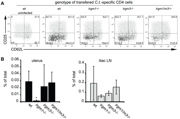
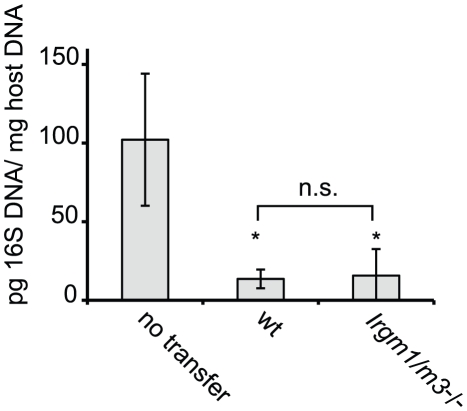
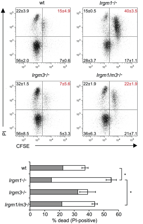
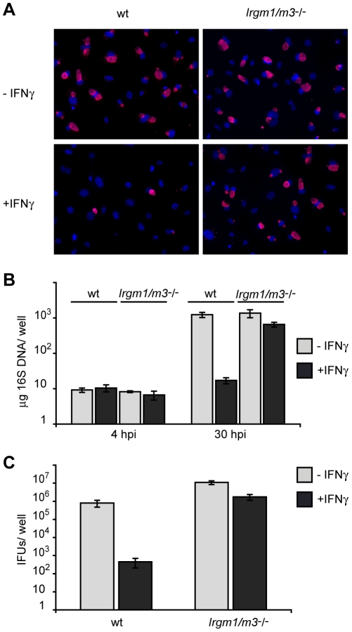
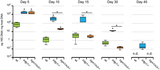
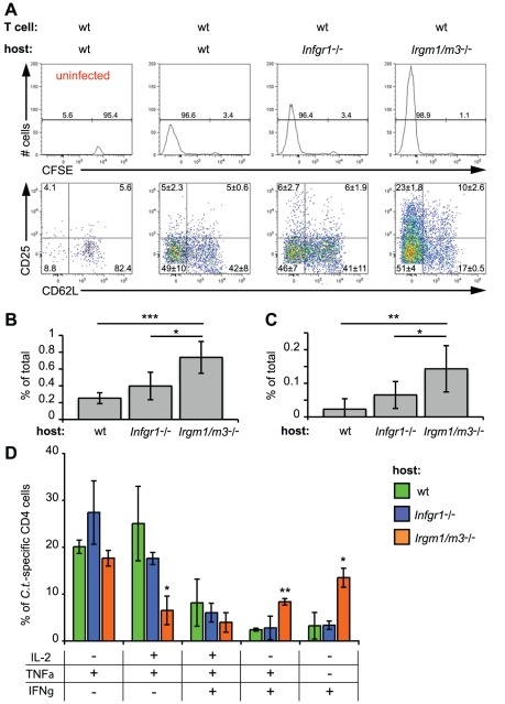
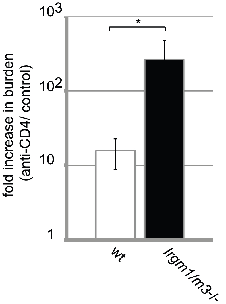
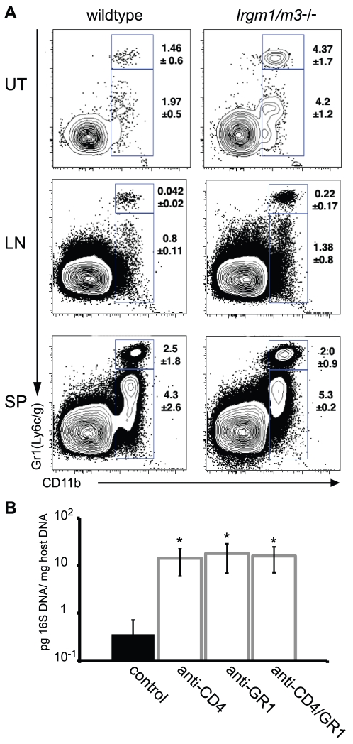
The obligate intracellular pathogen Chlamydia trachomatis is the most common cause of bacterial sexually transmitted diseases in the United States. In women C. trachomatis can establish persistent genital infections that lead to pelvic inflammatory disease and sterility. In contrast to natural infections in humans, experimentally induced infections with C. trachomatis in mice are rapidly cleared. The cytokine interferon-γ (IFNγ) plays a critical role in the clearance of C. trachomatis infections in mice. Because IFNγ induces an antimicrobial defense system in mice but not in humans that is composed of a large family of Immunity Related GTPases (IRGs), we questioned whether mice deficient in IRG immunity would develop persistent infections with C. trachomatis as observed in human patients. We found that IRG-deficient Irgm1/m3((-/-)) mice transiently develop high bacterial burden post intrauterine infection, but subsequently clear the infection more efficiently than wildtype mice. We show that the delayed but highly effective clearance of intrauterine C. trachomatis infections in Irgm1/m3((-/-)) mice is dependent on an exacerbated CD4(+) T cell response. These findings indicate that the absence of the predominant murine innate effector mechanism restricting C. trachomatis growth inside epithelial cells results in a compensatory adaptive immune response, which is at least in part driven by CD4(+) T cells and prevents the establishment of a persistent infection in mice.


Conflict of interest statement

The authors have declared that no competing interests exist.

Figures

Figure 1
Figure 1. Concomitant deletion of Irgm3 rescues the expansion defect of Irgm1 -/- CD4+ T cells.
Wildtype B6 mice were injected with naïve, NR1-transgene expressing T cells derived from mice with the indicated genotypes. The next day mice were challenged with 2 * 106 IFUs of C. trachomatis directly instilled into the uterus. On day 6 post-infection, flow cytometry was used to analyze cells from the uterus and draining lymph node (A). C. trachomatis-specific NR1 cells resident in the iliac lymph nodes were assessed for the expression of activation markers CD25 and CD62L. Total CD4+ cells are shown as gray dots, NR1 cells are shown as black dots. (B) The percentage of total live NR1 cells was calculated for uterus (left panel) and draining lymph node (right panel). Flow cytometry data are representative of three independent experiments. Statistical analysis was performed via Student's t test. *, p<0.05.
Figure 2
Figure 2. C. trachomatis-specific CD4+ T cells lacking Irgm1 and Irgm3 convey protection to an intrauterine infection with C. trachomatis.
Th1-skewed wildtype NR1 cells or Irgm1/m3 (-/-) NR1 cells were adoptively transferred into mice deficient for IFNγ production (Ifng -/- mice). The following day these mice were transcervically infected with 2 * 106 IFUs of C. trachomatis. Three days post-infection, uteri were taken from sacrificed mice and bacterial yield was quantified using qPCR. Data are representative of two independent experiments. Statistical significance relative to the no transfer group is indicated as *, p<0.05. n.s.  =  not significant.
Figure 3
Figure 3. Concomitant deletion of Irgm3 rescues the cell death phenotype of Irgm1 -/- CD4+ T cells.
Flow cytometry of CFSE-labeled wildtype, Irgm1 -/-, Irgm3 -/- and Irgm1/m3 (-/-) CD4+ T cells that had been activated with anti-CD3 and anti-CD28 antibodies in the presence of IL-2 for 72 hours and were subsequently stained for dead cells with propidium iodide. Percentages and standard deviations are given for each quadrant (upper panel). The total percentage of propidium iodide positive cells is shown for each genotype. The filled area of the bar indicates the fraction of CFSElow cells and the open area marks the fraction of CFSEhigh cells (lower panel). Data are representative of three independent experiments. Statistical significance for the percentages of total cell death is indicated as *, p<0.05.
Figure 4
Figure 4. Cells doubly deficient for Irgm1 and Irgm3 exert minimal IFNγ-induced cell-autonomous resistance to C. trachomatis.
IFNγ-stimulated and unstimulated MEFs derived from wildtype B6 or Irgm1/m3 (-/-) mice were infected with C. trachomatis at an MOI of 2. (A) Cell nuclei were stained with DAPI and C. trachomatis inclusions were labeled using an anti-Hsp70 antibody (B) Bacterial growth was quantified using qPCR at 4 and 30 hpi and (C) using an inclusion-forming unit (IFU) assay at 48 hpi. Data is representative of at least two independent experiments.
Figure 5
Figure 5. Irgm1/m3 (-/-) mice show delayed but efficient clearance of genital C. trachomatis infections.
B6 wildtype mice and the coisogenic knockout strains B6.Ifngr1 -/-, and B6.Irgm1/m3 (-/-) were transcervically infected with 2 * 106 IFUs of C. trachomatis. At the indicated days post-infection 3 to 4 mice of each genotype were sacrificed and bacterial burden in the uteri was quantified by qPCR. Data were combined from two independent experiments. Statistical analysis of box-and-whisker plots was done via Student's t test and significant difference relative to wildtype mice and between Ifngr1 -/-, and Irgm1/m3 (-/-) are indicated by *, p<0.05. The limit of detection was approximately 10−4 pg 16S DNA/µg host DNA.
Figure 6
Figure 6. The absence of Irgm1/3-dependent immunity results in an exacerbated CD4+ T cell response.
CFSE-labeled, wildtype NR1cells were adoptively transferred into recipient mice of the indicated genotypes. The mice were subsequently transcervically infected with 2 * 106 IFUs of C. trachomatis. On day 6 post-infection, flow cytometry was used to analyze cells from the uterus and draining lymph node. Representative data of four independent experiments are shown. (A) A total of 106 lymphocytes were collected in each sample and the absolute number of CD4+ CD90.1+ NR1 cells was analyzed for CFSE dye dilution (upper panel) and expression of the surface proteins CD25 and CD62L (lower panel). All cells derived from the draining lymph nodes were used for the analysis. The number of C. trachomatis-specific NR1 cells is shown as the percentage of total number of cells in the iliac lymph node (B) and the uterus (C). (D) NR1 cells harvested from the iliac lymph node were also restimulated for 5 h with PMA-ionomycin and assessed for intracellular cytokine staining. Statistical significance relative to wildtype recipient mice is shown. *, p<0.05; **, p<0.01; and ***, p<0.005.
Figure 7
Figure 7. Dependency on the CD4+ T cell response for clearing intrauterine C. trachomatis infections is increased in Irgm1/m3 (-/-) mice.
Groups of wildtype B6 and Irgm1/m3 (-/-) mice were treated with either ant-CD4 depleting or control antibodies as described in Materials and Methods. At 15 days post-infection mice were sacrificed and bacterial burden in the uteri was determined by qPCR. The fold increase in bacterial burden following treatment with anti-CD4 antibody compared to control antibody-treated mice is shown. The increase in burden in anti-CD4-treated Irgm1/m3 (-/-) mice was significantly higher than in wildtype mice (*, p<0.05). Data are representative of three independent experiments.
Figure 8
Figure 8. A neutrophil surge into the uterus of C. trachomatis-infected Irgm1/m3 (-/-) mice plays a role in immune clearance.
(A) Wildtype or Irgm1/m3 (-/-) mice were transcervically infected with C. trachomatis. Fifteen days later, the uterus (UT), iliac lymphnodes (LN), and spleens (SP) of these mice were assessed for lymphocyte populations via FACS analysis. Macrophage populations were gated via CD11b+GR1low, while neutrophil populations exhibited a CD11b+GR1high phenotype. Average percent of total is represented on the right of the gated population +/- standard deviation. Neutrophil populations in uteri and lymph nodes were statistically different between wildtype and Irgm1/m3 (-/-) mice (p<0.05). (B) Irgm1/m3 (-/-) mice were treated with depleting antibodies as described in Materials and Methods and 15 days post-infection bacterial burden in the spleen was measured by qPCR. Significant increases in bacterial burden in antibody-treated Irgm1/m3 (-/-) mice compared to mice treated with an isotype control antibody are indicated (*, p<0.05). The data are representative of two independent experiments.

References

    1. Belland R, Ojcius DM, Byrne GI. Chlamydia. Nat Rev Microbiol. 2004;2:530–531. - PubMed
    1. Resnikoff S, Pascolini D, Etya'ale D, Kocur I, Pararajasegaram R, et al. Global data on visual impairment in the year 2002. Bull World Health Organ. 2004;82:844–851. - PMC - PubMed
    1. World Health Organization. World Health Organization: Geneva; 2003. World Health Report, Shaping the future.
    1. Beatty WL, Morrison RP, Byrne GI. Persistent chlamydiae: from cell culture to a paradigm for chlamydial pathogenesis. Microbiol Rev. 1994;58:686–699. - PMC - PubMed
    1. Mpiga P, Ravaoarinoro M. Chlamydia trachomatis persistence: an update. Microbiol Res. 2006;161:9–19. - PubMed

Publication types