. 2011 Jun;7(6):e1002085.
doi: 10.1371/journal.ppat.1002085. Epub 2011 Jun 23.

The N-terminus of the RNA polymerase from infectious pancreatic necrosis virus is the determinant of genome attachment

Affiliations

The N-terminus of the RNA polymerase from infectious pancreatic necrosis virus is the determinant of genome attachment

Stephen C Graham et al. PLoS Pathog. 2011 Jun.

Abstract

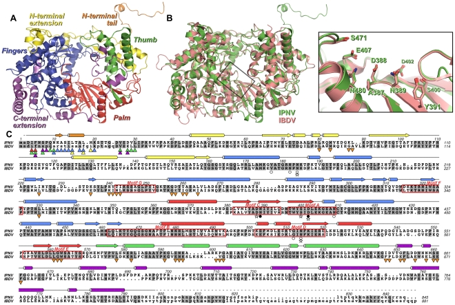
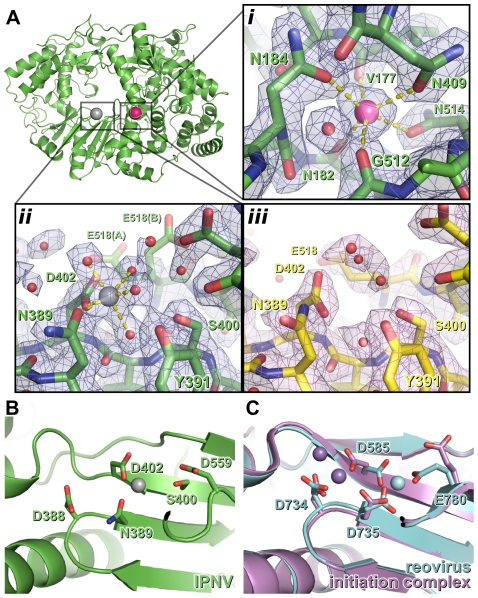
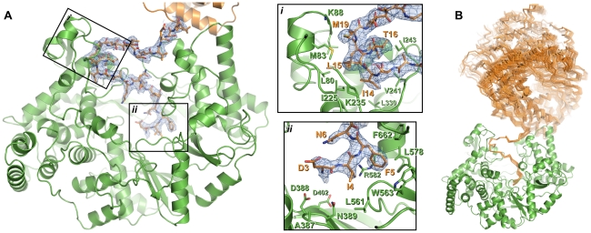
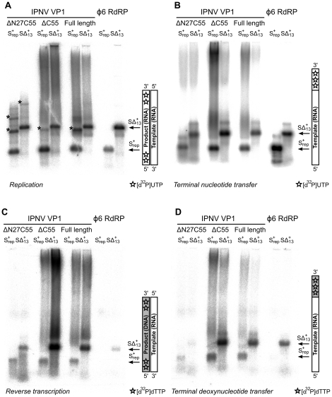
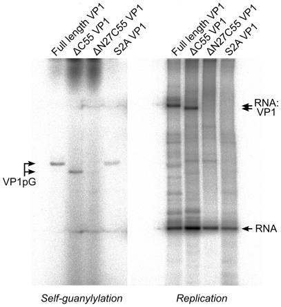
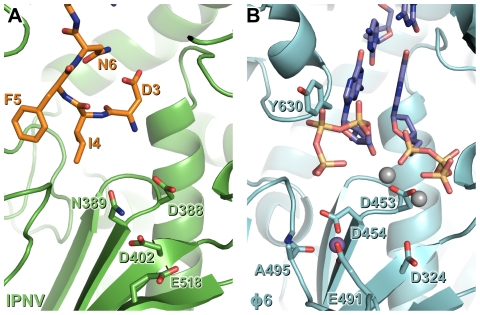
The RNA-dependent RNA polymerase VP1 of infectious pancreatic necrosis virus (IPNV) is a single polypeptide responsible for both viral RNA transcription and genome replication. Sequence analysis identifies IPNV VP1 as having an unusual active site topology. We have purified, crystallized and solved the structure of IPNV VP1 to 2.3 Å resolution in its apo form and at 2.2 Å resolution bound to the catalytically-activating metal magnesium. We find that recombinantly-expressed VP1 is highly active for RNA transcription and replication, yielding both free and polymerase-attached RNA products. IPNV VP1 also possesses terminal (deoxy)nucleotide transferase, RNA-dependent DNA polymerase (reverse transcriptase) and template-independent self-guanylylation activity. The N-terminus of VP1 interacts with the active-site cleft and we show that the N-terminal serine residue is required for formation of covalent RNA:polymerase complexes, providing a mechanism for the genesis of viral genome:polymerase complexes observed in vivo.


Conflict of interest statement

The authors have declared that no competing interests exist.

Figures

Figure 1
Figure 1. Structure of IPNV VP1.
(A) Cartoon representation of the structure of IPNV ΔC55 VP1, colored to show the canonical RdRP fingers (blue), thumb (green) and palm (red) domains, the N-terminal (yellow) and C-terminal (magenta) extensions to the canonical RdRP fold and the N-terminal tail (orange). (B) Superposition of VP1 from IPNV (green) and IBDV (pink; PDB ID 2PGG). The inset shows the superposed active sites, for clarity only selected side chains are shown. (C) Structure-based sequence alignment of IPNV and IBDV VP1. IPNV secondary structure is shown above the sequences, colored as in (A). Conserved residues are highlighted in grey, the characteristic polymerase motifs are boxed and residues not observed in the structures are in lower case. Triangles denote residues of the N-terminal tail (▴) or polymerase domain (▾) that interact with each other, the color of the triangle corresponding to the region of VP1 with which this residue interacts (colors being as in (A)). Stars denote residues mutated in this study, filled circles represent Mg2+-binding residues and empty circles denote K+-binding residues.
Figure 2
Figure 2. Metal binding of IPNV VP1.
(A) The structure of IPNV ΔC55 VP1 soaked with Mg2+ is shown in cartoon representation, rotated by approximately 180° around a vertical axis relative to the orientation in Figure 1. Mg2+ and K+ ions are shown as oversized grey and pink spheres, respectively. (i) K+ binding site in 2F O-F C electron density calculated from the final refined model (blue, 1.5 σ). (ii) Mg2+ binding site, electron density is as in (i). (iii) The active site of apo ΔC55 VP1 in 2F O-F C electron density calculated from the final refined model (magenta, 1.5 σ). (B) Mg2+ (grey) binds away from the ‘tip’ of the motif C catalytic β hairpin at the active site of IPNV VP1. View is rotated by approximately 45° around the horizontal axis compared with (A). (C) Binding of the Mn2+ at the active site of unliganded reovirus RdRP (cyan; PBB ID 1MWH) and of a reovirus RdRP initiation complex (purple; PBB ID 1N1H). View is as in (B). For clarity, in (B) and (C) only selected side chains are shown and NTPs are omitted.
Figure 3
Figure 3. Interaction of the N-terminal tail with the polymerase domain of VP1.
(A) The polymerase domain of VP1 is shown in cartoon representation (green), the N-terminal tail that interacts with the active site of the polymerase domain is shown as sticks (orange) and the polymerase domain from which this N-terminal tail extends is shown in cartoon representation (orange). 2F O-F C electron density calculated from the final refined model is shown in blue (1.0 σ); for clarity density is shown only within a 2 Å radius of N-terminal tail residues. Insets (i) and (ii) show close-up views of the interaction between the N-terminal tail and the polymerase domain. For clarity side chains are shown only for polymerase domain residues that interact with the N-terminal tail or lie at the active site, and only selected residues are labeled. (B) Orientation of the polymerase domain from which the N-terminal tail extends (orange Cα trace) relative to the polymerase domain to which the N-terminal tail binds (green cartoon). Sixteen independent views of the interacting polymerase domain (from three different crystal forms) are shown, all having been superposed upon residues 11–19 of a reference interaction.
Figure 4
Figure 4. Catalytic activities of recombinant IPNV VP1.
Autoradiography following agarose gel electrophoresis of [α32P]-UTP (A and B) or [α32P]-dTTP (C and D) labeled (A) de novo replication, (B) terminal nucleotide transfer, (C) reverse transcription and (D) terminal deoxynucleotide transfer reactions catalyzed by ΔN27C55, ΔC55 and full-length VP1 or the Φ6 RdRP. Note the high molecular weight smear present in reactions catalyzed by full-length or ΔC55 VP1, absent in reactions catalyzed by ΔN27C55 VP1, and the presence of discrete larger reaction products in (A) consistent with concatenation of product RNA chains (marked with *).
Figure 5
Figure 5. IPNV VP1 self-guanylylation and RNA∶polymerase complex formation.
Autoradiography following denaturing SDS PAGE of IPNV VP1 self-guanylylation (left panel) and de novo RNA polymerization using s+ rep template (300 nt ssRNA, right panel) in the presence of [α32P]-GTP. Full-length and ΔC55 VP1 self-guanylylate to form covalent VP1pG complexes (left panel) and form covalent RNA∶polymerase complexes migrating at approximately 180–200 kDa (right panel). ΔN27C55 VP1 lacks both these activities, while an S2A mutant of the full-length enzyme retains self-guanylylation activity but lacks the ability to form covalent RNA∶polymerase complexes.
Figure 6
Figure 6. N-terminal tail binding at the active site of IPNV VP1 resembles the nascent daughter strand of a viral polymerase initiation complex.
Mg2+ ions are shown in grey and Mn2+ ions in purple, and for clarity only selected side chains are shown. (A) IPNV VP1 (green) with residues from the N-terminal tail of an adjacent molecule in the crystal shown as sticks (orange). (B) Φ6 RdRP initiation complex (cyan; PDB ID 1HI0). The GTP molecules that will form the 5′ nucleotides of the nascent strand and the template 3′ nucleotides to which they Watson-Crick base pair are shown (blue).

References

    1. Bruenn JA. A structural and primary sequence comparison of the viral RNA-dependent RNA polymerases. Nucleic Acids Res. 2003;31:1821–1829. - PMC - PubMed
    1. Delarue M, Poch O, Tordo N, Moras D, Argos P. An attempt to unify the structure of polymerases. Protein Eng. 1990;3:461–467. - PubMed
    1. Gorbalenya AE, Pringle FM, Zeddam JL, Luke BT, Cameron CE, et al. The palm subdomain-based active site is internally permuted in viral RNA-dependent RNA polymerases of an ancient lineage. J Mol Biol. 2002;324:47–62. - PMC - PubMed
    1. Coulibaly F, Chevalier C, Gutsche I, Pous J, Navaza J, et al. The birnavirus crystal structure reveals structural relationships among icosahedral viruses. Cell. 2005;120:761–772. - PubMed
    1. Muller H, Islam MR, Raue R. Research on infectious bursal disease–the past, the present and the future. Vet Microbiol. 2003;97:153–165. - PubMed

Publication types