. 2011 Jun;7(6):e1002097.
doi: 10.1371/journal.pgen.1002097. Epub 2011 Jun 23.

Interferon regulatory factor 8 regulates pathways for antigen presentation in myeloid cells and during tuberculosis

Affiliations

Interferon regulatory factor 8 regulates pathways for antigen presentation in myeloid cells and during tuberculosis

Jean-François Marquis et al. PLoS Genet. 2011 Jun.

Abstract

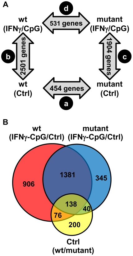
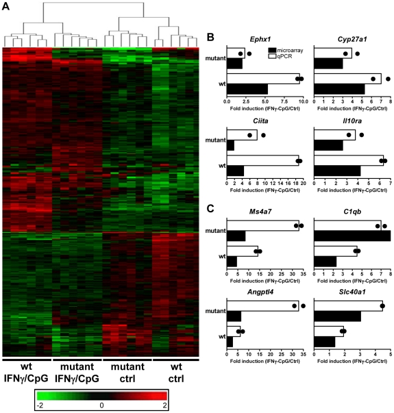
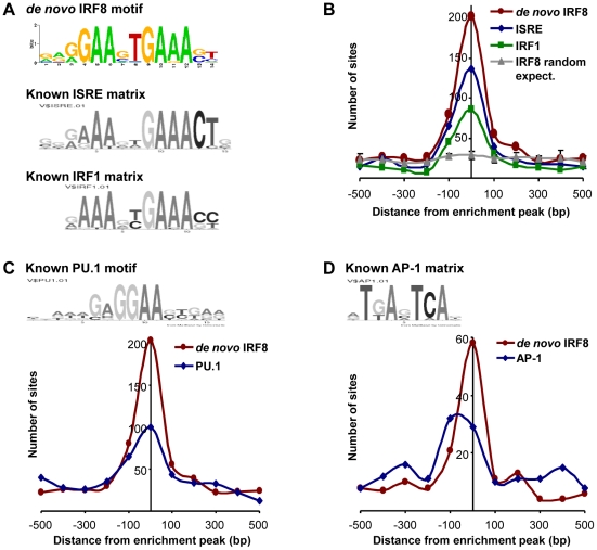
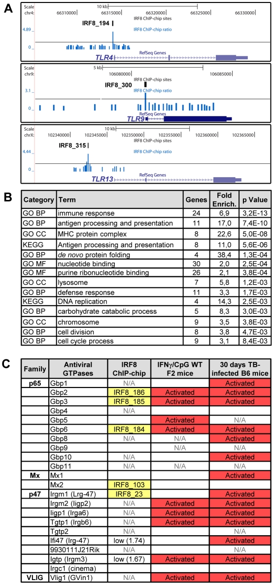
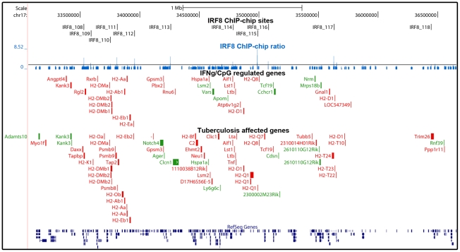
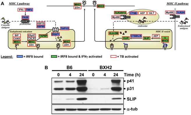
IRF8 (Interferon Regulatory Factor 8) plays an important role in defenses against intracellular pathogens, including several aspects of myeloid cells function. It is required for ontogeny and maturation of macrophages and dendritic cells, for activation of anti-microbial defenses, and for production of the Th1-polarizing cytokine interleukin-12 (IL-12) in response to interferon gamma (IFNγ) and protection against infection with Mycobacterium tuberculosis. The transcriptional programs and cellular pathways that are regulated by IRF8 in response to IFNγ and that are important for defenses against M. tuberculosis are poorly understood. These were investigated by transcript profiling and chromatin immunoprecipitation on microarrays (ChIP-chip). Studies in primary macrophages identified 368 genes that are regulated by IRF8 in response to IFNγ/CpG and that behave as stably segregating expression signatures (eQTLs) in F2 mice fixed for a wild-type or mutant allele at IRF8. A total of 319 IRF8 binding sites were identified on promoters genome-wide (ChIP-chip) in macrophages treated with IFNγ/CpG, defining a functional G/AGAAnTGAAA motif. An analysis of the genes bearing a functional IRF8 binding site, and showing regulation by IFNγ/CpG in macrophages and/or in M. tuberculosis-infected lungs, revealed a striking enrichment for the pathways of antigen processing and presentation, including multiple structural and enzymatic components of the Class I and Class II MHC (major histocompatibility complex) antigen presentation machinery. Also significantly enriched as IRF8 targets are the group of endomembrane- and phagosome-associated small GTPases of the IRG (immunity-related GTPases) and GBP (guanylate binding proteins) families. These results identify IRF8 as a key regulator of early response pathways in myeloid cells, including phagosome maturation, antigen processing, and antigen presentation by myeloid cells.


Conflict of interest statement

The authors have declared that no competing interests exist.

Figures

Figure 1
Figure 1. Pairwise analysis of transcriptional responses of wt and IRF8 mutant BMDMs at basal level and following exposure to IFNγ/CpG.
BMDMs RNA was obtained from individual wt and IRF8 mutant F2 mice either prior to (unstimulated control) or 3 hrs following stimulation with IFNγ/CpG (6 samples per experimental group; 24 samples in total), and hybridized to microarrays. The 3 hrs IFNγ/CpG-stimulated macrophage cultures were initially primed with IFNγ (50 U/ml) for 18 hrs. (A) A closed-loop strategy was applied to monitor differences in transcript abundance between the four experimental groups; (comparison a) mutant control versus wt control; (comparison b) wt stimulated versus wt control; (comparison c) mutant stimulated versus mutant control; (comparison d) mutant stimulated versus wt stimulated. The numbers of significantly modulated transcripts identified for each single pairwise comparison are indicated within the gray arrows. (B) A Venn diagram analysis of the pairwise comparisons a, b, and c revealed considerable overlaps in the lists of transcripts which level of expression is affected by the IRF8 alleles and the IFNγ/CpG stimulation.
Figure 2
Figure 2. Transcriptional programs elicited by IFNγ/CpG exposure in wt and IRF8 mutant F2 mice.
BMDMs RNA was obtained from individual wt and IRF8 mutant F2 mice either prior to (unstimulated control) or 3 hrs following stimulation with IFNγ/CpG (6 samples per experimental group; 24 samples in total), and hybridized to microarrays. (A) By using a 2×2 interaction Anova analysis, 368 genes were recognized to be significantly differentially modulated by IRF8 in response to IFNγ/CpG exposure between the wt and IRF8 mutant cells. The expression profiles are ordered by hierarchical clustering; the genes, illustrated by their specific signal intensities (Log2 scale) are displayed as rows and individual mouse samples/conditions as columns. Red coloring signifies high level of expression; green coloring denotes low level of expression. The dendrogram illustrates the clustering of the samples according to expression pattern similarities. RNA samples (6 per experimental group) used for this transcriptional profiling analysis were pooled and used for qPCR validation. Ephx1, Cyp27a1, Ciita, and Il10ra were selected as genes positively affected by the presence of functional IRF8 following IFNγ/CpG exposure (B), while Ms4a7, C1qb, Angptl4, and Slc40a1 were selected as genes negatively affected by the presence of a functional IRF8 following IFNγ/CpG exposure (C). The ratios of expression (IFNγ/CpG-stimulated versus unstimulated control), represented by fold induction, were calculated for the wt and IRF8 mutant mice separately (white bars), and compared to the corresponding microarray results (black bars). The black dots indicate the qPCR values obtained for each replicate. The microarray results were statistically significant according to Anova analysis (t test p value of 0.05 and a fold-change cutoff of 1.5X). Hprt was used to standardize the mRNA levels of target genes for qPCR.
Figure 3
Figure 3. Transcription factor binding motif analyses on IRF8 ChIP-chip binding sites.
IRF8 chromatin immunoprecipitated from IFNγ activated macrophages was hybridized to Agilent promoter tiling array (ChIP-chip). After normalization and statistical analysis, we identified 319 IRF8 binding sites with a threshold of 2 fold enrichment and p value≤0.001 relative to a non-specific control antibody ChIP. (A) De novo binding motif analysis was carried out on 500 bp sequence flanking the IRF8 sites. The MEME algorithm returned an IRF-like motif as the top motif with a high score. The known ISRE and IRF1 motifs are from the Genomatix MatBase database. (B) The 319 IRF8 binding sites were queried for the IRF8 de novo binding motif and for the ISRE and IRF1 motif shown in A. The position of the in silico found sites was plotted relative to the ChIP-chip IRF8 binding peak. They all cluster over the peak center, whereas there is no IRF8 motif enrichment in a set of randomly chosen sequences. (C) The PU.1 (ETS family member) motif shows a clear colocalization with the IRF8 de novo motif, as reported by the in silico analysis. (D) The AP-1 motif is also enriched.
Figure 4
Figure 4. IRF8 regulates important functions of APC cells.
(A) IRF8 ChIP-chip binding profiles extracted from the UCSC genome browser for Toll-like receptors (Tlr4,9,13); Black rectangles represent the significant binding peak and the blue bars correspond to ChIP-chip binding ratios. (B) Gene ontology (GO) and KEGG pathways analysis of the genes regulated by IFNγ-CpG treatment in F2 wt mice and having an IRF8 binding site in their proximity. The IRF8 targets genes are implicated in immune response, nucleotide binding and antigen processing and presentation. (C) Antiviral GTPases gene regulation summary. Association of known antiviral GTPases with IRF8 binding sites, IFNγ/CpG regulation in wt F2 mice on Illumina Mouse WG-6 v2.0 Expression BeadChip arrays and regulation after M. tuberculosis infection in B6 mice on Affymetrix oligonucleotides chips (Mouse Genome 430 2.0 array). N/A indicates that this was not assessed by the microarrays.
Figure 5
Figure 5. Enrichment of IRF8 targets within the boundaries of the MHC locus.
IRF8 ChIP-chip binding profiles extracted from the UCSC genome browser for the MHC locus. The black rectangles represent the significant binding peak and the blue bars correspond to ChIP-chip binding ratios. Genes regulated by IFNγ/CpG and/or M. tuberculosis infection are represented by red (activation) and green (repression) boxes.
Figure 6
Figure 6. The IRF8 binding sites are strongly associated with antigen presenting cells function.
(A) Schematic representation of the “antigen processing and presentation” pathway. Adapted from the KEGG pathway database , and current literature . Genes implicated in Class I and Class II MHC antigen processing and presentation were annotated for IRF8 binding (blue box), for activated by IFNγ-CpG (green box), and for activation by infection with M. tuberculosis (red outline). These results suggest that IRF8 is a key regulator of all steps of the antigen processing and presentation pathway. (B) Validation of Cd74 (Ii) at protein level. BMDMs total proteins were obtained from wt (B6) and IRF8 mutant (BXH2) mice either prior to (unstimulated control) or following stimulation with IFNγ/CpG (4 hrs and 24 hrs post-stimulation), separated in 10% SDS-PAGE and probed with In-1 mAb. The migration of intact p41 and p31 Ii, as well as SLIP Ii fragment (∼12-kD) is indicated. α-tubulin was used as a constitutively expressed internal control to normalize the protein levels.

References

    1. Flannagan RS, Cosio G, Grinstein S. Antimicrobial mechanisms of phagocytes and bacterial evasion strategies. Nat Rev Microbiol. 2009;7:355–366. - PubMed
    1. Casanova JL, Abel L. Human genetics of infectious diseases: a unified theory. EMBO J. 2007;26:915–922. - PMC - PubMed
    1. Hill AV. Aspects of genetic susceptibility to human infectious diseases. Annu Rev Genet. 2006;40:469–486. - PubMed
    1. Lipoldova M, Demant P. Genetic susceptibility to infectious disease: lessons from mouse models of leishmaniasis. Nat Rev Genet. 2006;7:294–305. - PubMed
    1. Vidal SM, Malo D, Marquis JF, Gros P. Forward genetic dissection of immunity to infection in the mouse. Annu Rev Immunol. 2008;26:81–132. - PubMed

Publication types

MeSH terms

Substances