Pneumopericardium as a complication of pericardiocentesis
- PMID: 21731571
- PMCID: PMC3116108
- DOI: 10.4070/kcj.2011.41.5.280
Pneumopericardium as a complication of pericardiocentesis
Abstract
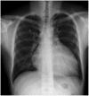
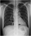
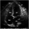
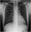
Pneumopericardium is a rare complication of pericardiocentesis, occurring either as a result of direct pleuro-pericardial communication or a leaky drainage system. Air-fluid level surrounding the heart shadow within the pericardium on a chest X-ray is an early observation at diagnosis. This clinical measurement and process is variable, depending on the hemodynamic status of the patient. The development of a cardiac tamponade is a serious complication, necessitating prompt recognition and treatment. We recently observed a case of pneumopericardium after a therapeutic pericardiocentesis in a 20-year-old man with tuberculous pericardial effusion.
Keywords: Pericardiocentesis; Pneumopericardium.
Conflict of interest statement
The authors have no financial conflicts of interest.
Figures


References
-
- Arroyo M, Soberman JE. Adenosine deaminase in the diagnosis of tuberculous pericardial effusion. Am J Med Sci. 2008;335:227–229. - PubMed
-
- Mayosi BM, Burgess LJ, Doubell AF. Tuberculous pericarditis. Circulation. 2005;112:3608–3616. - PubMed
-
- Park WH, Jun JE, Park MH. Echocardiographic observation in 50 cases of pericardial effusion. Korean Circ J. 1982;12:135–143.
LinkOut - more resources
Full Text Sources

