. 2011;6(6):e20945.
doi: 10.1371/journal.pone.0020945. Epub 2011 Jun 22.

Simvastatin prevents dopaminergic neurodegeneration in experimental parkinsonian models: the association with anti-inflammatory responses

Affiliations

Simvastatin prevents dopaminergic neurodegeneration in experimental parkinsonian models: the association with anti-inflammatory responses

Junqiang Yan et al. PLoS One. 2011.

Abstract

Background: In addition to their original applications to lowering cholesterol, statins display multiple neuroprotective effects. N-methyl-D-aspartate (NMDA) receptors interact closely with the dopaminergic system and are strongly implicated in therapeutic paradigms of Parkinson's disease (PD). This study aims to investigate how simvastatin impacts on experimental parkinsonian models via regulating NMDA receptors.

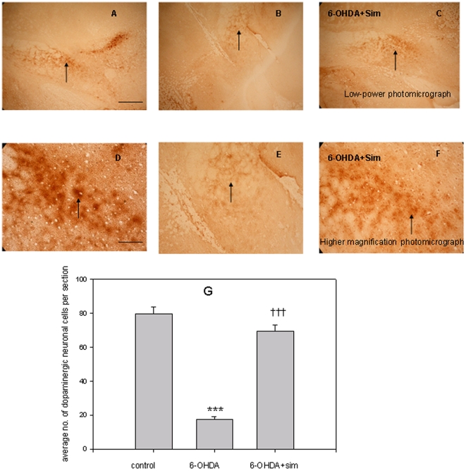
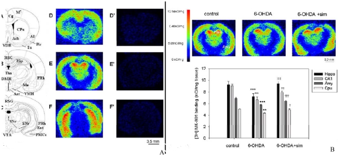
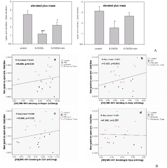
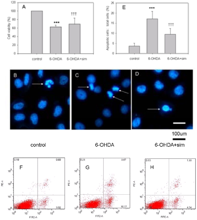
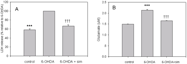
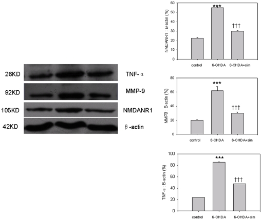
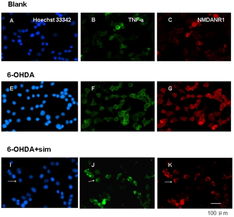
Methodology/principal findings: Regional changes in NMDA receptors in the rat brain and anxiolytic-like activity were examined after unilateral medial forebrain bundle lesion by 6-hydroxydopamine via a 3-week administration of simvastatin. NMDA receptor alterations in the post-mortem rat brain were detected by [³H]MK-801(Dizocilpine) binding autoradiography. 6-hydroxydopamine treated PC12 was applied to investigate the neuroprotection of simvastatin, the association with NMDA receptors, and the anti-inflammation. 6-hydroxydopamine induced anxiety and the downregulation of NMDA receptors in the hippocampus, CA1(Cornu Ammonis 1 Area), amygdala and caudate putamen was observed in 6-OHDA(6-hydroxydopamine) lesioned rats whereas simvastatin significantly ameliorated the anxiety-like activity and restored the expression of NMDA receptors in examined brain regions. Significant positive correlations were identified between anxiolytic-like activity and the restoration of expression of NMDA receptors in the hippocampus, amygdala and CA1 following simvastatin administration. Simvastatin exerted neuroprotection in 6-hydroxydopamine-lesioned rat brain and 6-hydroxydopamine treated PC12, partially by regulating NMDA receptors, MMP9 (matrix metalloproteinase-9), and TNF-a (tumour necrosis factor-alpha).

Conclusions/significance: Our results provide strong evidence that NMDA receptor modulation after simvastatin treatment could partially explain its anxiolytic-like activity and anti-inflammatory mechanisms in experimental parkinsonian models. These findings contribute to a better understanding of the critical roles of simvastatin in treating PD via NMDA receptors.


Conflict of interest statement

Competing Interests: The authors have declared that no competing interests exist.

Figures

Figure 1
Figure 1. Effects of 6-OHDA lesion and simvastatin on TH immunohistochemistry staining in the SNpc.
Figs. A, B, C shows TH staining in low-power photomicrograph in the SNpc of unlesioned, 6-OHDA-lesioned, and 6-OHDA-lesioned with simvastatin treatment groups, respectively. Bar = 450 µm. Figs. D, E, F shows TH staining at higher magnification photomicrograph in the SNpc of unlesioned, 6-OHDA-lesioned, and 6-OHDA-lesioned with simvastatin treated groups, respectively. Bar = 120 µm. Fig. 1G represents the average number of TH-positive dopaminergic neurons in the SNpc of unlesioned (control), 6-OHDA lesioned, and 6-OHDA lesioned with simvastatin treatment groups. The values represent mean ±SEM, n = 6–8. ***p<0.001, 6-OHDA group versus control group; ††† p<0.001, 6-OHDA+simvastatin group versus 6-OHDA group.
Figure 2
Figure 2
2A.[3H] MK-801 autoradiography depicts the expression of NMDA receptors in the rat brain. The maps of A, B and C are adopted from a rat brain atlas indicating the levels where the [3H]MK-801 binding density was measured. Autoradiographs (D, E, F) and (D', E', F') depict the expression of [3H]MK-801 binding and non-specific [3H]MK-801 binding at different rostro-caudal coronal levels of the rat brain. 2B. Typical autoradiographs depict the expression of NMDA receptors in the hippocampus (Hipp) and amygdala (Amy) among control, 6-OHDA-lesioned rats, and 6-OHDA lesioned rats that also received simvastatin treatment. The bar chart shows the effects of chronic simvastatin treatment on [3H]MK-801 binding in the different groups of rat brain regions. Note: Units of measurement are in nCi/mg tissue. Data are means ± SEM. Asterisks indicate significant differences from control group (saline) and cross indicates significant differences between 6-OHDA rats and 6-OHDA with simvastatin-treated rats (n = 6–8, **p<0.01; ***p<0.001; †p<0.05; ††p<0.01; †††p<0.001, one-way ANOVA followed by Tukey's test).
Figure 3
Figure 3
3A.Simvastatin ameliorates the anxiety of 6-OHDA rats in the EPM test. The graph shows the ratio of time spent in the open arms to total time and the ratio of open arm entries to total entries in the EPM. The parameters are expressed as a percentage of time spent in the open arms to the total time and open arm entries to total entries in the EPM. The values represent mean ± SEM, n = 6–8. †p<0.05, 6-OHDA group versus 6-OHDA+simvastatin group for open arm duration; ***p<0.001, 6-OHDA group versus control group for open arm duration; *p<0.05, 6-OHDA group versus control group for open entires. 3B. Correlations between duration in the open arm of EPM and [3H]MK-801 binding density in brain regions. A significant positive correlation was identified between the [3H]MK-801 binding density in the hippocampus (r = 0.485 Pearson's correlation, p = 0.026), amygdala (r = 0.622, p = 0.003), CA1 (r = 0.638, p = 0.002), respectively, and the time spent in the open arm of the EPM.
Figure 4
Figure 4. Simvastatin protected PC12 cells against 6-OHDA neurotoxicity.
The MTT value in the 6-OHDA treated group was significantly reduced as compared with controls (***p<0.001, 6-OHDA vs controls, n = 9; Fig. 4A), but simvastatin upregulated this reduction (†††p<0.001, 6-OHDA vs 6-OHDA+sim, n = 9; Fig. 4A). Intact nuclei (blue Hoechst 33342 staining) and condensed/fragmented nuclei (bright blue Hoechst 33342 staining) were considered to be live and apoptotic cells, respectively (Fig. 4B, C, D). The exposure of the PC12 cultures to 6-OHDA (100 uM, 24 h) significantly increased the number of apoptotic cells by 4.75 times compared with controls (***p<0.001, 6-OHDA vs controls; Fig. 4E); however, simvastatin incubation significantly reduced this increase in the number of apoptotic cells (†††p<0.001, 6-OHDA vs 6-OHDA+sim; Fig. 4E; Bar = 100 µm). Apoptotic cells were further verified by flow cytometry analysis. The result showed that 6-OHDA induced profound apoptosis (4.59±0.9% vs 14.97±1.25%, controls vs 6-OHDA, p<0.01, n = 5; Fig. 4F and 6G) but simvastatin incubation attenuated this apoptotic death (14.97±1.25% vs 6.09±0.64%, 6-OHDA vs 6-OHDA+sim, p<0.01, n = 5; Fig. 4G and 4H). All the results are expressed as mean ± standard error of the mean.
Figure 5
Figure 5. Simvastatin reduced 6-OHDA-induced LDH and glutamate.
LDH in 6-OHDA incubated PC12 increased by 1.74 times compared with controls (***p<0.001, 6-OHDA vs controls, n = 9; Fig. 5A), but simvastatin incubation abolished this elevation (†††p<0.001, 6-OHDA vs 6-OHDA+sim, n = 9; Fig. 5A). In 6-OHDA incubated PC12, glutamate was increased by 1.43 times compared with controls (2.138±0.03 µm vs 1.49±0.01 µm, 6-OHDA vs controls, ***p<0.001, n = 9; Fig. 5B), but simvastatin treatment abolished this elevation (2.138±0.03 µm vs 1.64±0.01 µm, 6-OHDA vs 6-OHDA+sim, †††p<0.001, n = 9; Fig. 5B). All of the results are expressed as mean ± standard error of the mean.
Figure 6
Figure 6. Simvastatin reduced 6-OHDA medicated elevations of NMDANR1 receptors, TNF-a, and MMP9.
6-OHDA incubation pronouncedly increased the NR1 receptors compared with controls (***p<0.001, 6-OHDA vs controls, n = 6–9); while this elevation was significantly abolished following simvastatin treatment (†††p<0.001, 6-OHDA vs 6-OHDA+sim, n = 6–9). Compared with controls, 6-OHDA produced significant increases in the total amount of TNF-a and MMP9 (***p<0.001, 6-OHDA vs controls, n = 6–9); while these increases were prevented by simvastatin treatment (†††p<0.001, 6-OHDA vs 6-OHDA + sim, n = 6–9). All the results are expressed as mean ± standard error of the mean.
Figure 7
Figure 7. 6-OHDA increased synaptic cluster density and number of clusters NR1 receptors and TNF-a, and the upregulation was abolished after simvastatin treatment.
Arrows in I, J, K indicate nuclear, TNF-a, and NR1, respectively. PC12 cultures double-labeled for NR1 (red, C,G,K) and TNF-a (green, B,F,J); Hoechst 33342 indicates nuclear staining (blue, A, E, I). 6-OHDA treatment significantly increased the density of NR1 (G) and TNF-a clusters (F), and the elevated density was abolished by simvastatin treatment (K, J for NR1 and TNF-a, respectively). A significant difference in the density of NR1 and TNF-a was observed among control, 6-OHDA, and 6-OHDA+sim groups (p<0.05, control vs 6-OHDA; p<0.05, 6-OHDA vs 6-OHDA+sim; n = 9–12; Student's t test). All the results are expressed as means ± standard error of the mean. Scale bars: 100 µm.

References

    1. Becker C, Jick SS, Meier CR. Use of statins and the risk of Parkinson's disease: a retrospective case-control study in the UK. Drug Saf. 2008;31(5):399–407. - PubMed
    1. Wang Q, Yan J, Chen X, Li J, Yang Y, et al. Statins: Multiple neuroprotective mechanisms in neurodegenerative diseases. Exp Neurol. 2010 In Press. - PubMed
    1. Janssen WG, Vissavajjhala P, Andrews G, Moran T, Hof PR, et al. Cellular and synaptic distribution of NR2A and NR2B in macaque monkey and rat hippocampus as visualized with subunit-specific monoclonal antibodies. Exp Neurol. 2005;191(Suppl 1):S28–44. - PubMed
    1. Nilsson A, Eriksson M, Muly EC, Akesson E, Samuelsson EB, et al. Analysis of NR3A receptor subunits in human native NMDA receptors. Brain Res. 2007;1186:102–112. - PubMed
    1. Yu SP, Sensi SL, Canzoniero LM, Buisson A, Choi DW. Membrane-delimited modulation of NMDA currents by metabotropic glutamate receptor subtypes 1/5 in cultured mouse cortical neurons. J Physiol. 1997;499(Pt 3):721–732. - PMC - PubMed

Publication types

MeSH terms