. 2011;6(6):e21216.
doi: 10.1371/journal.pone.0021216. Epub 2011 Jun 22.

Daidzein prevents the increase in CD4+CD28null T cells and B lymphopoesis in ovariectomized mice: a key mechanism for anti-osteoclastogenic effect

Affiliations

Daidzein prevents the increase in CD4+CD28null T cells and B lymphopoesis in ovariectomized mice: a key mechanism for anti-osteoclastogenic effect

Abdul Malik Tyagi et al. PLoS One. 2011.

Abstract

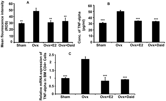
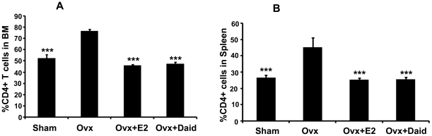
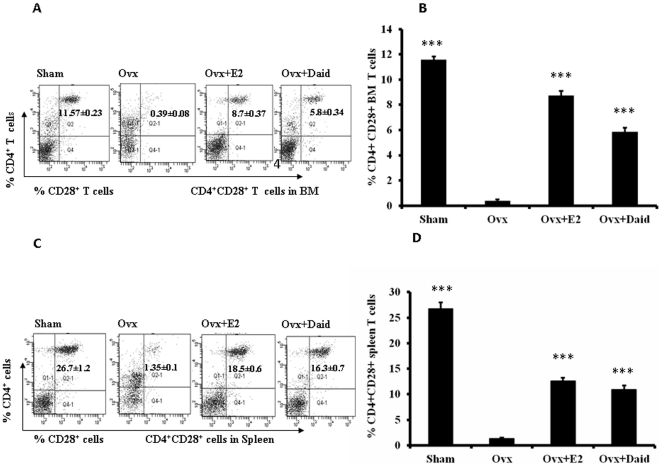
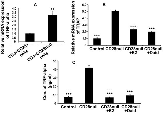
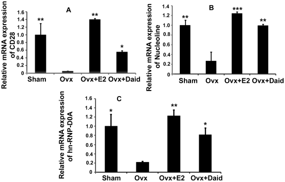
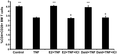
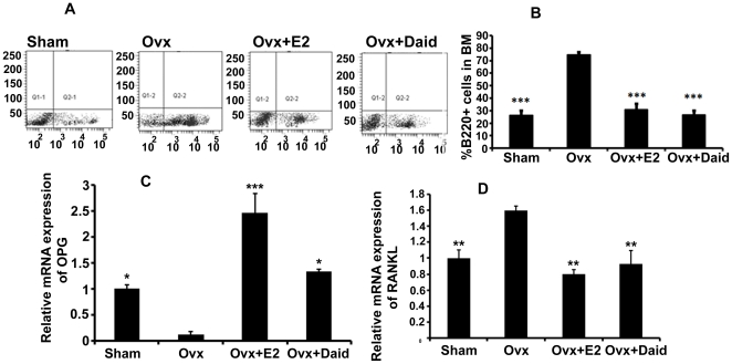
Estrogen deficiency leads to an upregulation of TNF-α producing T cells and B-lymphopoesis which augments osteoclastogenesis. Estrogen deficiency also increases the population of premature senescent CD4⁺ CD28null T cells which secrete a higher amount of TNF-α thus leading to enhanced osteoclastogenesis. Isoflavonoids like daidzein and genistein are found mostly in soybeans, legumes, and peas. These share structural similarity with 17β-stradiol (E2) and have osteoprotective role. This study explores the effect of daidzein (Daid) on the proliferation of TNF-α producing T cells, premature senescent T cells and B cell lymphopoesis under estrogen deficient conditions. For this study adult Balb/c mice were treated with Daid at 10 mg/kg body weight dose by oral gavage daily post ovariectomy (Ovx). After six weeks animals were autopsied and bone marrow and spleen cells were collected for FACS analysis. Blood serum was collected for ELISA. It was observed that Ovx mice treated with Daid for six weeks show reduction in Ovx induced expansion of CD4⁺ T cells in bone marrow and spleen when analysed by flow cytometry. Estrogen deficiency led to increased prevalence of TNF-α secreting CD4⁺CD28null T cells, however, treatment with Daid increased the percentage of CD4⁺CD28⁺ T cells. Co-culture of CD4⁺CD28null T cells and bone marrow resulted in enhanced osteoclastogenesis as evident by increased tartarate resistant acid phosphatase (TRAP) expression, an osteoclast marker. However, treatment with Daid resulted in reduced osteoclastogenesis in CD4⁺CD28null T cells and bone marrow cell co-culture. Daid also regulated B lymphopoesis and decreased mRNA levels of RANKL in B220⁺ cells. Taken together, we propose that one of the mechanisms by which Daid prevents bone loss is by reversing the detrimental immune changes as a result of estrogen deficiency.


Conflict of interest statement

Competing Interests: The authors have declared that no competing interests exist.

Figures

Figure 1
Figure 1. Daid treatment inhibits Ovx-induced oxidative stress in CD4+ T cells and TNF-α production by these cells.
(A) Cellular ROS measurement was performed by incubating CD4+ T cells with DCF-DA followed by FACS analysis. (B) Circulating TNF-α levels were measured in various groups by ELISA. (C) TNF-α mRNA levels in the BM CD4+ T cells were measured in various groups by qPCR. N = 10 mice/group; data are presented as mean ± SEM; ** P<0.01 and *** P<0.001 compared with Ovx+vehicle group.
Figure 2
Figure 2. Daid treatment significantly decreased Ovx-induced increases in CD4+ T cell subsets in the BM and spleen.
(A) CD4+ T cells in BM and (B) CD4+ T cells in spleen were quantified by flow cytometry as described in Materials and Methods. N = 10 mice/group; data are presented as mean ± SEM; *** P<0.001 compared with Ovx+vehicle group.
Figure 3
Figure 3. Daid treatment prevented Ovx-induced loss of CD28 on the BM as well as in spleen CD4+ T cells.
(A) Representative image (B) CD4+CD28+ T cells in BM, (C) Representative image (D) CD4+CD28+ T cells in spleen of Sham, Ovx, Daid and E2 were measured by flow cytometry as described in Materials and Methods. N = 10 mice/group; data are presented as mean ± SEM; *** P<0.001 compared with Ovx+vehicle group.
Figure 4
Figure 4. Daidzein inhibits increased osteoclastogenesis in co-culture of bone marrow cells and TNF-α secreting CD4+CD28null T cells.
(A) TRAP mRNA expression by qPCR in co-culture of BM cells and CD4+CD28null T cells. N = 3 mice/group; data are presented as mean ± SEM; *** P<0.001 compared with untreated co-culture of CD4+CD28null T cells with BM cells. (B) TNF-α levels were measured in the co-culture by ELISA. N = 3 mice/group; data are presented as mean ± SEM; *** P<0.001 compared with untreated co-culture of CD4+CD28null T cells with BM cells.
Figure 5
Figure 5. Daid treatment increased CD28, nucleolin and hnRNP-D0A mRNA levels assessed by qPCR in the BM T cells of Ovx mice.
mRNA levels of CD28, nucleolin and hnRNP-D0A in CD4+ T cells (A–C). All data are the mean ± SD of three independent experiments; n = 3 mice/group; * P<0.05, ** P<0.01, *** P<0.001 compared with Ovx+vehicle group.
Figure 6
Figure 6. Daid abrogates TNF-α mediated CD28 downregulation in the BM CD4+ T cells.
(A) BM CD4+ T cells (5×10⁵ cells/well) were seeded in 24-well plates. For ICI 182,780 (ICI) treatment group, the cells were pre-treated with ICI for 30 min. Various treatments as shown were given to CD4+ T cells for 24 h [E2-10−9 M, Daid −10−8 M, TNF-α−10.0 ng/ml]. At the end of incubation, cells were stained with antibodies against CD3, CD4 and CD28 and subjected to flow cytometry as described in Materials and Methods. Data are presented as mean ± SEM from 3 independent experiments; *** P<0.001 compared with TNF-α treated cells and c P<0.001 compared between Daid+TNF-α and Daid+TNF-α+ICI treated cells; E2+TNF-α and E2+TNF-α+ICI treated cells.
Figure 7
Figure 7. Daid treatment significantly decreased Ovx-induced increases in B220+ cells in BM.
(A and B) B220+ cells in BM were quantified by flow cytometry as described in Materials and Methods. (C) OPG mRNA levels in the BM B220+ cells were measured in various groups by qPCR. (D) RANKL mRNA levels in the BM B220+ cells were measured in various groups by qPCR. N = 10 mice/group; data are presented as mean ± SEM; * P<0.05, ** P<0.01, *** P<0.001 compared with Ovx+vehicle group.

References

    1. Roggia C, Gao Y, Cenci S, Weitzmann MN, Toraldo G, et al. Up-regulation of TNF-producing T cells in the bone marrow: a key mechanism by which estrogen deficiency induces bone loss in vivo. Proc Natl Acad Sci U S A. 2001;98:13960–13965. - PMC - PubMed
    1. Weitzmann MN, Pacifici R. The role of T lymphocytes in bone metabolism. Immunol Rev. 2005;208:154–168. - PubMed
    1. Pacifici R. T cells and post menopausal osteoporosis in murine models. Arthritis Res Ther. 2007;9:102. - PMC - PubMed
    1. D'Amelio P, Grimaldi A, Di Bella S, Brianza SZ, Cristofaro MA, et al. Estrogen deficiency increases osteoclastogenesis up-regulating T cells activity: a key mechanism in osteoporosis. Bone. 2008;43:92–100. - PubMed
    1. Vallejo AN, Weyand CM, Goronzy JJ. T cell senescense: a culprit of immune abnormalities in chronic inflammation and persistent infection. Trends in Molecular Medicine. 2004;10:119–124. - PubMed

Publication types

MeSH terms