. 2011;6(6):e21386.
doi: 10.1371/journal.pone.0021386. Epub 2011 Jun 24.

T regulatory cells are markers of disease activity in multiple sclerosis patients

Affiliations

T regulatory cells are markers of disease activity in multiple sclerosis patients

Dacia Dalla Libera et al. PLoS One. 2011.

Abstract

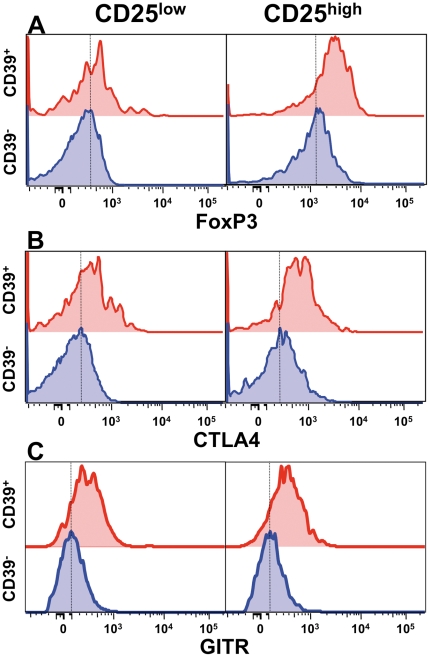
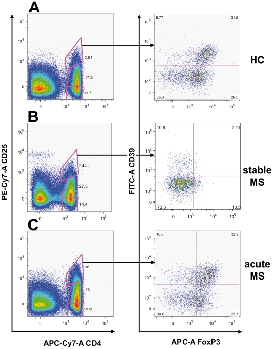
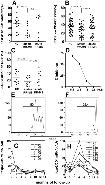
FoxP3⁺ Treg cells are believed to play a role in the occurrence of autoimmunity and in the determination of clinical recurrences. Contradictory reports are, however, available describing frequency and function of Treg cells during autoimmune diseases. We examined, by both polychromatic flow cytometry, and real-time RT-PCR, several Treg markers in peripheral blood mononuclear cells from patients with multiple sclerosis (MS), an autoimmune disease affecting the central nervous system. We found that Tregs, as defined by CD25, CD39, FoxP3, CTLA4, and GITR expression, were significantly decreased in stable MS patients as compared to healthy donors, but, surprisingly, restored to normal levels during an acute clinical attack. We conclude that Treg cells are not involved in causing clinical relapses, but rather react to inflammation in the attempt to restore homeostasis.


Conflict of interest statement

Competing Interests: The authors have declared that no competing interests exist.

Figures

Figure 1
Figure 1. CD4+CD25highCD39+ T cells express increased levels of Treg markers.
FoxP3 (A), CTLA4 (B) and GITR (C) expression, as measured by flow cytometry, are increased in CD4+CD39+, as compared to CD4+CD39 T cells, especially in the CD25high compartment.
Figure 2
Figure 2. Treg markers are up-regulated in RR-MS patients experiencing clinical relapses.
A–E. Clinically relapsing RR-MS patients displayed increased PBMC mRNA levels for CD25 (A), CTLA-4 (B), GITR (C), CD39 (D), and foxp3 (E). We also found significantly lower levels of Treg markers mRNA in stable RR-MS patients as compared to healthy controls (HC). Values are expressed as arbitrary units (AU). P values are indicated (Mann-Whitney).
Figure 3
Figure 3. FoxP3+CD39+ Treg cells are increased during acute MS.
Scatter plots from three representative subjects, one healthy donor (HC; A), a stable MS patients (B), and a patient experiencing a clinical re-exacerbation of MS (C) are shown, indicating that the FoxP3/CD39 double positive T cell population (A, right panel) in the CD25high gate dramatically decreases during stable MS (B, right panel) and is restored during an acute attack (C, right panel).
Figure 4
Figure 4. Treg cells are increased during acute MS.
The percentage of FoxP3+ cells in the CD4+CD25high gate is shown in (A) in healthy controls (HC; n = 14), stable MS (n = 10), and acute MS (n = 8). The percentage of CD39+ cells in the CD4+CD25high gate is shown in (B) in HC (n = 44), stable MS (n = 31), and acute MS (n = 32), and, similarly, the percentage of FoxP3+/CD39+ cells in the CD4+CD25high gate is displayed in (C) in HC (n = 13), stable MS (n = 12), and acute MS (n = 11). Again, while Treg cells, as defined by these markers, were significantly decreased in stable MS patients and restored during an acute attack. Lines represent median values, and P values are indicated where significant (Mann-Whitney). CD4+CD25highCD39+ regulatory T cells from an acute MS patient suppress T responder cell proliferation (E) in a dose dependent way (F), as measured by CFSE dilution assay. Plots represent CFSE-labeled T responder cell proliferation in absence and presence of regulatory T cell (5∶1) (E, F). A representative experiment among three is shown. Treatment-free RR-MS patients (n = 15) were followed longitudinally every two months for 14 months (G–H), and divided according to the occurrence of clinical relapses during the follow-up in stable (G), or relapsing (H) patients. PBMC foxp3 mRNA values, normalized on CD4 and GAPDH mRNA, and expressed as arbitrary units (AU) are plotted.

References

    1. Kasper LH, Haque A, Haque S. Regulatory mechanisms of the immune system in multiple sclerosis. T regulatory cells: turned on to turn off. J Neurol. 2007;254(Suppl 1):I10–I14.
    1. Kohm AP, Carpentier PA, Anger HA, Miller SD. Cutting edge: CD4+CD25+ regulatory T cells suppress antigen-specific autoreactive immune responses and central nervous system inflammation during active experimental autoimmune encephalomyelitis. J Immunol. 2002;169:4712–4716. - PubMed
    1. Reddy J, Illes Z, Zhang X, Encinas J, Pyrdol J, et al. Myelin proteolipid protein-specific CD4+CD25+ regulatory cells mediate genetic resistance to experimental autoimmune encephalomyelitis. Proc Natl Acad Sci U S A. 2004;101:15434–15439. - PMC - PubMed
    1. Huan J, Culbertson N, Spencer L, Bartholomew R, Burrows GG, et al. Decreased FOXP3 levels in multiple sclerosis patients. J Neurosci Res. 2005;81:45–52. - PubMed
    1. Putheti P, Pettersson A, Soderstrom M, Link H, Huang YM. Circulating CD4+CD25+ T regulatory cells are not altered in multiple sclerosis and unaffected by disease-modulating drugs. J Clin Immunol. 2004;24:155–161. - PubMed

Publication types