. 2011;6(6):e21473.
doi: 10.1371/journal.pone.0021473. Epub 2011 Jun 24.

A novel missense mutation in the connexin30 causes nonsyndromic hearing loss

Affiliations

A novel missense mutation in the connexin30 causes nonsyndromic hearing loss

Wen-Hung Wang et al. PLoS One. 2011.

Abstract

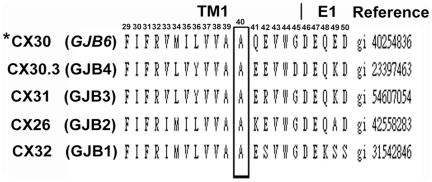
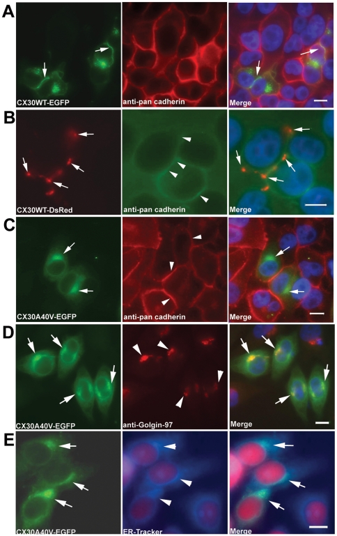
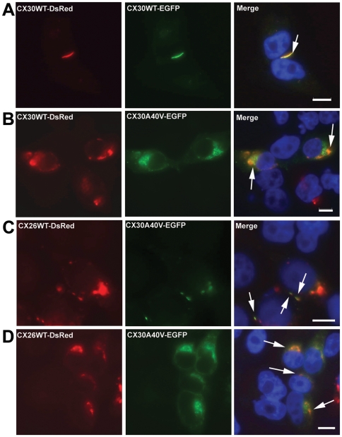
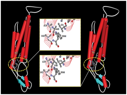
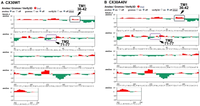
Dysfunctional gap junctions caused by GJB2 (CX26) and GJB6 (CX30) mutations are implicated in nearly half of nonsyndromic hearing loss cases. A recent study identified a heterozygous mutation, c.119C>T (p.A40V), in the GJB6 gene of patients with nonsyndromic hearing loss. However, the functional role of the mutation in hearing loss remains unclear. In this study, analyses of cell biology indicated that a p.A40V missense mutation of CX30 causes CX30 protein accumulation in the Golgi body rather than in the cytoplasmic membrane. The tet-on protein expression system was used for further study of mutant proteins in CX30 and CX30A40V co-expressions and in CX26 and CX30A40V co-expressions. The p.A40V missense mutation exerted a dominant negative effect on both normal CX30 and CX26, which impaired gap junction formation. Moreover, computer-assisted modeling suggested that this p.A40V mutation affects the intra molecular interaction in the hydrophobic core of Trp44, which significantly alters the efficiency of gap junction formation. These findings suggest that the p.A40V mutation in CX30 causes autosomal-dominant nonsyndromic hearing loss. These data provide a novel molecular explanation for the role of GJB6 in hearing loss.


Conflict of interest statement

Competing Interests: The authors have declared that no competing interests exist.

Figures

Figure 1
Figure 1. Alignment of amino acid sequences of the TM1 domain of CX30 proteins in members of the human b “Insert>Symbol”-group of CX family.
The p.A40 residue, which was mutated in this study, is indicated in bold type. This alanine (A) at codon 40 is identical among members of human b “Insert>Symbol”-group of CX family. Human CX30 is indicated by an asterisk (*).
Figure 2
Figure 2. Analysis of CX30WT and CX30A40V expression in HeLa cells transiently transfected by immunocytochemistry.
Fluorescence microscopy results for CX30WT-EGFP (A) and CX30WT-DsRed (B) HeLa cells showing expression of CX30 fusion protein in the plasma membranes using pan-cadherin antibody. The CX30A40V-EGFP (C) transfected HeLa cells show impaired trafficking and concentration near the nucleus. Intercellular localization of mutant CX30A40V protein. Photomicrographs of HeLa cells transiently transfected with CX30A40V-EGFP cDNA and immunostained for use as markers of Golgi apparatus (D) and ER (E). The staining results for mutant CX30A40V showed substantial co-localization in the Golgi apparatus and moderate co-localization with ER marker. Cells were counterstained with DAPI to highlight the nuclei. Arrows indicate CX30 protein localization. Scale bars: 10 m“Insert>Symbol”m.
Figure 3
Figure 3. Co-expression of mutant proteins with CX30WT and CX26WT revealed by tet-on protein expression system.
(A) Tet-on HeLa cells co-expressing CX30WT-DsRed and CX30WT-EGFP. Co-localization of the two proteins is visible at the plasma membrane. (B) Tet-on HeLa cells co-expressing CX30WT-DsRed and CX30A40V-EGFP. Co-localization of the two proteins is visible near the nucleus regions. (C) Tet-on HeLa cells co-expressing CX30WT-DsRed and Cx26WT-EGFP. Co-localization of the two proteins is visible at the plasma membrane. (D) Tet-on HeLa cells co-expressing CX26WT-DsRed and CX30A40V-EGFP. Co-localization of the two proteins is visible near the nucleus. Arrows indicate co-expressed proteins. Cells were counterstained with DAPI to highlight the nuclei. Scale bars: 10 m“Insert>Symbol”m.
Figure 4
Figure 4. Bioinformatic estimation of three-dimensional (3D) structure of wildtype and p.A40V CX30 protein.
Comparison of protein secondary structure architectures reveals the intra-molecular interactions between wild type (A) and A40V mutation (B) in monomeric human CX30. Secondary structures appear as red solid cylinders (α-helices), blue arrows (β-sheets) and white loops (turns). The yellow boxes indicate the close-up views of intra-molecular interactions focus on the extracellular part (E1) of the transmembrane region (TM2) in a similar orientation as in ball and stick representation. The highly conserved residues in this region, including Ala39, Ala40, Val43 and Ile74, contributed to the hydrophobic core around Trp44, which stabilized the Cx30 intra-molecular structure.
Figure 5
Figure 5. Plot outputs of Anolea mean force potential reported from SWISS-MODEL workspace.
The graphs present the computer modeling results for wild-type (A) and p.A40V mutations (B) of the CX30 protein. Relative quality was evaluated by target-template similarity and modeling procedure. Non-local environments of each residue in the human Cx30 gene model (x-axis) were based on the template gene human Cx26 protein and on the percentage of energy change in all pairwise non-local interaction energy values between the template and modeling structure (represented by y-axis). Negative scores are plotted downwards and upwards to further validate the model by indicating the favorable and unfavorable energy environments, respectively. The arrows and boxes indicate that the two conserved regions contributing to the hydrophobic core around the key amino acid Trp44 were residues 36–42 located in the transmembrane 1 (TM1) region and residues 71–77 located in the TM2 region of the human Cx30 proteins, respectively.

References

    1. Petit C. Genes responsible for human hereditary deafness: symphony of a thousand. Nature Genet. 1996;14:385–391. - PubMed
    1. Apps SA, Rankin WA, Kurmis AP. Connexin26 mutations in autosomal recessive deafness disorders: A review. Int J Audiol. 2007;46:75–81. - PubMed
    1. Morton NE. Genetic epidemiogy of hearing impairment. Ann NY Acad Sci. 1991;630:16–31. - PubMed
    1. Pallares-Ruiz N, Blanchet P, Mondain M, Claustres M, Roux AF. A large deletion including most of GJB6 in recessive nonsyndromic deafness: a digenic effect? Eur J Hum Genet. 2002;10:72–76. - PubMed
    1. Beyer EC, Berthoud VM. Harris A, Locke D, editors. The family of connexin gene. Connexin: A Guide. 2009. pp. 3–26. Humana Press, Springer, USA.

Publication types

Supplementary concepts