

Immunohistochemical assessment of intrinsic and extrinsic markers of hypoxia in reproductive tissue: differential expression of HIF1α and HIF2α in rat oviduct and endometrium
- PMID: 21732047
- PMCID: PMC3136703
- DOI: 10.1007/s10735-011-9338-2
Immunohistochemical assessment of intrinsic and extrinsic markers of hypoxia in reproductive tissue: differential expression of HIF1α and HIF2α in rat oviduct and endometrium
Abstract
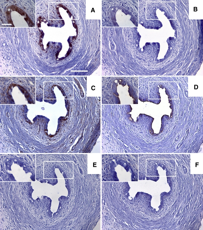
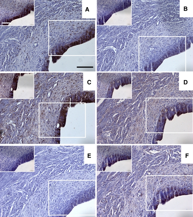
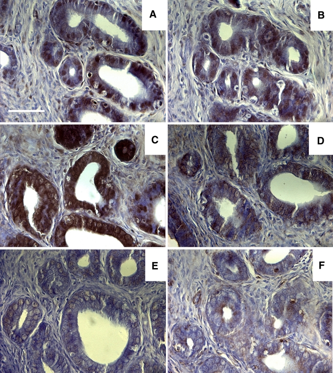
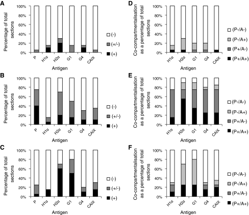
Hypoxia is thought to be critical in regulating physiological processes within the female reproductive system, including ovulation, composition of the fluid in the oviductal/uterine lumens and ovarian follicle development. This study examined the localisation of exogenous (pimonidazole) and endogenous [hypoxia inducible factor 1α and 2α (HIF1α, -2α), glucose transporter type 1 (GLUT1) and carbonic anhydrase 9 (CAIX)] hypoxia-related antigens within the oviduct and uterus of the rat reproductive tract. The extent to which each endogenous antigen co-compartmentalised with pimonidazole was also assessed. Female Wistar Furth rats (n = 10) were injected intraperitoneally with pimonidazole (60 mg/kg) 1 h prior to death. Reproductive tissues were removed immediately following death and fixed in 4% paraformaldehyde before being embedded in paraffin. Serial sections were cut (6-7 μm thick) and antigens of interest identified using standard immunohistochemical procedures. The mucosal epithelia of the ampulla, isthmus and uterus were immunopositive for pimonidazole in most sections. Co-compartmentalisation of pimonidazole with HIF1α was only expressed in the mucosa of the uterus whilst co-compartmentalisation with HIF2α was observed in the mucosa of the ampulla, isthmus and uterus. Both GLUT1 and CAIX were co-compartmentalised with pimonidazole in mucosa of the isthmus and uterus. This study confirms that mucosal regions of the rat oviduct and uterus frequently experience severe hypoxia and there are compartment specific variations in expression of endogenous hypoxia-related antigens, including the HIF isoforms. The latter observation may relate to target gene specificity of HIF isoforms or perhaps HIF2α's responsiveness to non-hypoxic stimuli such as hypoglycaemia independently of HIF1α.
Figures


Similar articles
-
The immunolocalization of HIF-2α, GLUT1 and CAIX in porcine oviduct during the estrous cycle.Anat Rec (Hoboken). 2023 Jan;306(1):176-186. doi: 10.1002/ar.25014. Epub 2022 Jun 23. Anat Rec (Hoboken). 2023. PMID: 35684983 Free PMC article.
-
Hypoxia inducible factor (HIf1alpha and HIF2alpha) and carbonic anhydrase 9 (CA9) expression and response of head-neck cancer to hypofractionated and accelerated radiotherapy.Int J Radiat Biol. 2008 Jan;84(1):47-52. doi: 10.1080/09553000701616114. Int J Radiat Biol. 2008. PMID: 17852557
-
Comparison between pimonidazole binding, oxygen electrode measurements, and expression of endogenous hypoxia markers in cancer of the uterine cervix.Cytometry B Clin Cytom. 2006 Mar;70(2):45-55. doi: 10.1002/cyto.b.20086. Cytometry B Clin Cytom. 2006. PMID: 16456867
-
Progress on hypoxia-inducible factor-3: Its structure, gene regulation and biological function (Review).Mol Med Rep. 2015 Aug;12(2):2411-6. doi: 10.3892/mmr.2015.3689. Epub 2015 Apr 27. Mol Med Rep. 2015. PMID: 25936862 Review.
-
Multiplicity of hypoxia-inducible transcription factors and their connection to the circadian clock in the zebrafish.Physiol Biochem Zool. 2015 Mar-Apr;88(2):146-57. doi: 10.1086/679751. Epub 2015 Jan 14. Physiol Biochem Zool. 2015. PMID: 25730270 Review.
Cited by
-
The immunolocalization of HIF-2α, GLUT1 and CAIX in porcine oviduct during the estrous cycle.Anat Rec (Hoboken). 2023 Jan;306(1):176-186. doi: 10.1002/ar.25014. Epub 2022 Jun 23. Anat Rec (Hoboken). 2023. PMID: 35684983 Free PMC article.
-
Tumor oxygenation and cancer therapy-then and now.Br J Radiol. 2019 Jan;92(1093):20170955. doi: 10.1259/bjr.20170955. Epub 2018 Mar 14. Br J Radiol. 2019. PMID: 29513032 Free PMC article. Review.
-
Hypoxia acts as an environmental cue for the human tissue-resident memory T cell differentiation program.JCI Insight. 2021 May 24;6(10):e138970. doi: 10.1172/jci.insight.138970. JCI Insight. 2021. PMID: 34027895 Free PMC article.
-
Oviductal Oxygen Homeostasis in Patients with Uterine Myoma: Correlation between Hypoxia and Telocytes.Int J Mol Sci. 2022 May 31;23(11):6155. doi: 10.3390/ijms23116155. Int J Mol Sci. 2022. PMID: 35682833 Free PMC article.
-
Curcumin ameliorates insulin signalling pathway in brain of Alzheimer's disease transgenic mice.Int J Immunopathol Pharmacol. 2016 Dec;29(4):734-741. doi: 10.1177/0394632016659494. Epub 2016 Jul 27. Int J Immunopathol Pharmacol. 2016. PMID: 27466310 Free PMC article.
References
-
- Chae KS, Kang MJ, Lee JH, Ryu BK, Lee MG, Her NG, Ha TK, Han J, Kim YK, Chi SG (2011) Opposite functions of HIF-alpha isoforms in VEGF induction by TGF-beta1 under non-hypoxic conditions. Oncogene 20(10):1213–1228 - PubMed
-
- Elvert G, Kappel A, Heidenreich R, Englmeier U, Lanz S, Acker T, Rauter M, Plate K, Sieweke M, Breier G, Flamme I. Cooperative interaction of hypoxia-inducible factor-2alpha (HIF-2alpha) and Ets-1 in the transcriptional activation of vascular endothelial growth factor receptor-2 (Flk-1) J Biol Chem. 2003;278(9):7520–7530. doi: 10.1074/jbc.M211298200. - DOI - PubMed
Publication types
MeSH terms
Substances
Grants and funding
LinkOut - more resources
Full Text Sources
Miscellaneous
