. 2012 Apr;227(4):1776-85.
doi: 10.1002/jcp.22911.

Requirements for ion and solute transport, and pH regulation during enamel maturation

Affiliations

Requirements for ion and solute transport, and pH regulation during enamel maturation

Rodrigo S Lacruz et al. J Cell Physiol. 2012 Apr.

Abstract

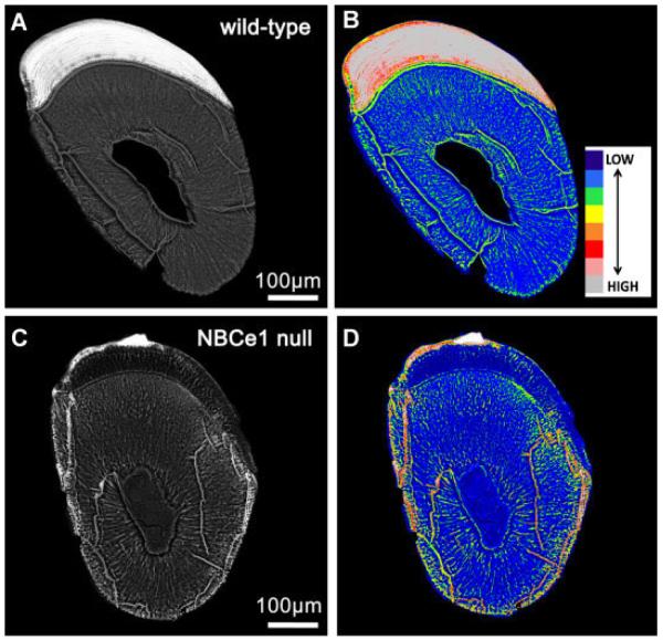
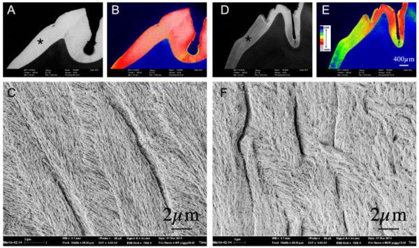
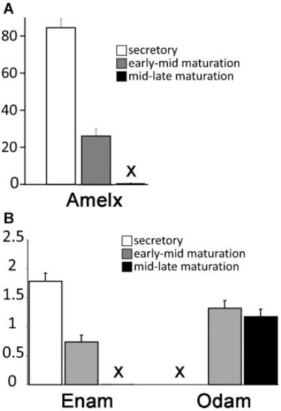
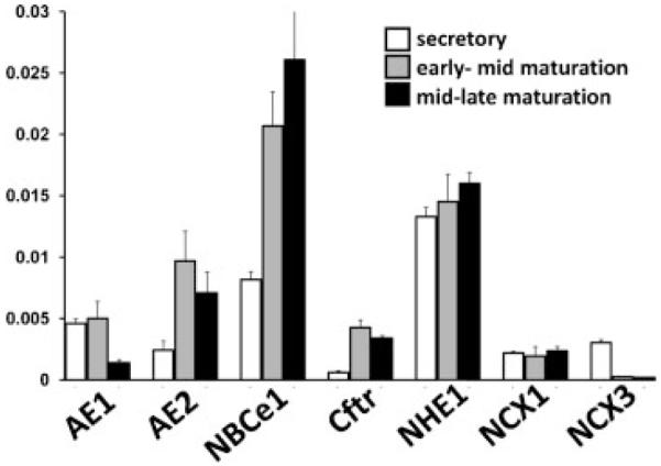
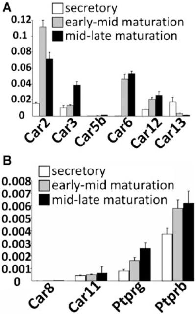
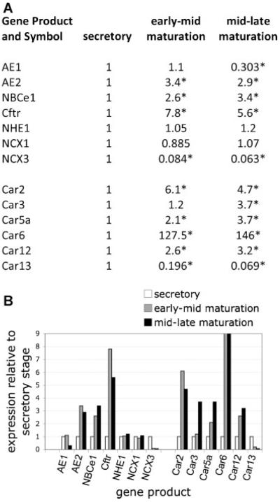
Transcellular bicarbonate transport is suspected to be an important pathway used by ameloblasts to regulate extracellular pH and support crystal growth during enamel maturation. Proteins that play a role in amelogenesis include members of the ABC transporters (SLC gene family and CFTR). A number of carbonic anhydrases (CAs) have also been identified. The defined functions of these genes are likely interlinked during enamel mineralization. The purpose of this study is to quantify relative mRNA levels of individual SLC, Cftr, and CAs in enamel cells obtained from secretory and maturation stages on rat incisors. We also present novel data on the enamel phenotypes for two animal models, a mutant porcine (CFTR-ΔF508) and the NBCe1-null mouse. Our data show that two SLCs (AE2 and NBCe1), Cftr, and Car2, Car3, Car6, and Car12 are all significantly up-regulated at the onset of the maturation stage of amelogenesis when compared to the secretory stage. The remaining SLCs and CA gene transcripts showed negligible expression or no significant change in expression from secretory to maturation stages. The enamel of CFTR-ΔF508 adult pigs was hypomineralized and showed abnormal crystal growth. NBCe1-null mice enamel was structurally defective and had a marked decrease in mineral content relative to wild-type. These data demonstrate the importance of many non-matrix proteins to amelogenesis and that the expression levels of multiple genes regulating extracellular pH are modulated during enamel maturation in response to an increased need for pH buffering during hydroxyapatite crystal growth.


Figures

Fig. 1
Fig. 1
Anatomical landmarks guiding the dissection and collection of secretory, early-mid maturation, and mid-late maturation enamel organ cells. The protocol described here has been adapted from the study of Smith and Nanci (1989) for our sample of rats weighing between 170 and 190 g. The study of Smith and Nanci (1989) used a molar reference line (black line apically from M1, and extending perpendicular to the labial aspect of the mandible) to obtain enamel organ cells (largely ameloblasts) from secretory (between “A” and “a”), early maturation (between “B” and “b”) and from late maturation (between “C” and “c”). In our study, the black star marks the position of the end of secretory stage in rats of 170–190 g, about 3 mm apically from the molar reference line. An area of 1 mm incisal to “a” was not collected to avoid possible contamination from post-secretory or transition cells.
Fig. 2
Fig. 2
BSE-SEM imaging of wild-type (A,B) and NBCe1-null (C,D) mice. Incisor teeth from 14-day-old NBCe1 null mice (Parts C and D) and an age-matched wild-type littermate (Parts A and B) were imaged in BSE-SEM using identical imaging parameters for both, which are defined in the text. Cross-sections are from an erupted, fully mature, region of the tooth. The enamel of the NBCe1-null mice is clearly hypomineralized. This is also evidenced by the color-coded images (Parts B andD), which illustrated marked differences in enamel mineralization for the NBCe1-null mice. Scale bars included.
Fig. 3
Fig. 3
FE-SEM imaging. The lower incisors of 15-day-old NBCe1 −/− mice and wild-type littermates were dissected out, air-dried, and mechanically fractured through the mature end (incisal) of the crown using a blade. Specimens were mounted on SEM stubs and carbon-coated for 6 sec to be imaged by FE-SEM. Parts A and B correspond to the enamel of a wild-type mouse, showing normal prisms and clear rod–interrod boundaries. The NBCe1 −/− mice (Parts C and D) lacked normal prismatic structure and present abnormally packed crystals. The EDJ has been highlighted in the NBCe1 −/− animal (dashed line in Part C). Scale bars included.
Fig. 4
Fig. 4
BSE-SEM and FE-SEM imaging of the permanent maxillary left first molar of a 6-month-old wild-type domestic pig (Parts A–C) compared toa1 2-month-old cystic fibrosis (Cftr-DF508) pig (PartsD–F).Low magnification BSE-SEM images of WT and Cftr-DF508 molars are provided in Parts A and D, respectively, both images of which were acquired under identical signal intensity settings. Scale for both is included in Part D. These images were subject to an 8-bin color look-up-table (Parts B and E) for visual comparison to represent differences in mineralization density between samples (see color code insert: wherein cool colors—blue, green, yellow denote lower mineralization density and hot colors—gray, pink, red represent higher mineralization densities). Asterisks on Parts A and D indicate areas shown in Parts C and F imaged at higher magnification (FW = 20 μm) by FE-SEM. Parts C and F represent WT and Cftr-DF508 molars, respectively, showing abnormal crystal growth in the latter as imaged in secondary electron emission mode.
Fig. 5
Fig. 5
Transcript analysis for Amelx (Part A), and Enam and Odam (Part B). The (X) in Parts A and B indicate that the measurements are present but negligible, with no appreciable error bars. The mRNA transcript levels were normalized to those of β-actin. Amelx and Enam expression clearly diminish at the onset of maturation, becoming almost completely absent as maturation progresses. The opposite is observed for Odam. These patterns are entirely consistent with reports on the activity levels of these genes using other methods. Therefore, we conclude that the dissected cells of the enamel organ analyzed in this study are well suited to investigate the expression levels of SLC genes and carbonic anhydrases.
Fig. 6
Fig. 6
Transcript analysis for AE1, AE2, NBCe1, Cftr, NHE1, NCX1 and NCX3. Note that the relative levels of AE1, AE2, NBCe1, Cftr, and NHE1 are of two orders of magnitude less than seen for Enam and Odam (Fig. 5). The mRNA transcript levels were normalized to those of β-actin.
Fig. 7
Fig. 7
Transcript analysis for selected carbonic anhydrases (Part A) and carbonic anhydrase-related proteins (Part B). Note that the relative levels of Car2, Car3, Car6, and Car12 are one order of magnitude less than seen for Enam and Odam (Fig. 5). While levels for Car5b (Part A), and the CA-RPs (Car8, Car11, Ptprg, and Ptprb) were recorded, the levels were low and negligible, as were the transcript levels for Car1, Car4, Car5a, Car7, Car9, Car14, and Car15 (data not included). The mRNA transcript levels were normalized to those of β-actin.
Fig. 8
Fig. 8
Relative levels of enamel epithelial gene up-regulation at early-mid and mid-late maturation stages of amelogenesis when compared to secretory stage amelogenesis, as determined by qPCR. For the selected gene transcripts, levels of both maturation stages were normalized against the level detected during secretory stage. Fold up-regulation is shown, as is the level of down-regulation. Data are presented numerically (Part A) and also as a graph (Part B). A cut-off fold value was used for Car6 in order to illustrate changes in other gene transcripts. Asterisks in Part A indicate statistically significant differences (P < 0.05), two-tailed Student’s t-test comparing secretory versus early-mid maturation or secretory to mid-late maturation stage amelogenesis.

References

    1. Aramaki S, Yoshida I, Yoshino M, Kondo M, Sato Y, Noda K, Jo R, Okue A, Sai N, Yamashita F. Carbonic anhydrase II deficiency in three unrelated Japanese patients. J Inherit Metab Dis. 1993;16:982–990. - PubMed
    1. Arquitt CK, Boyd C, Wright JT. Cystic fibrosis transmembrane regulator gene (CFTR) is associated with abnormal enamel formation. J Dent Res. 2002;81:492–496. - PubMed
    1. Atar M, Korperich EJ. Systemic disorders and their influence on the development of dental hard tissues: A literature review. J Dent. 2010;38:296–306. - PubMed
    1. Awad M, Al-Ashwal AA, Sakati N, Al-Abbad AA, Bin-Abbas BS. Long-term follow up of carbonic anhydrase II deficiency syndrome. Saudi Med J. 2002;23:25–29. - PubMed
    1. Azevedo TD, Feijo GC, Bezerra AC. Presence of developmental defects of enamel in cystic fibrosis patients. J Dent Child (Chic) 2006;73:159–163. - PubMed

Publication types

MeSH terms

LinkOut - more resources