. 2011 Feb;29(2):206-16.
doi: 10.1002/stem.566.

Osteoblasts derived from induced pluripotent stem cells form calcified structures in scaffolds both in vitro and in vivo

Affiliations

Osteoblasts derived from induced pluripotent stem cells form calcified structures in scaffolds both in vitro and in vivo

Ganna Bilousova et al. Stem Cells. 2011 Feb.

Abstract

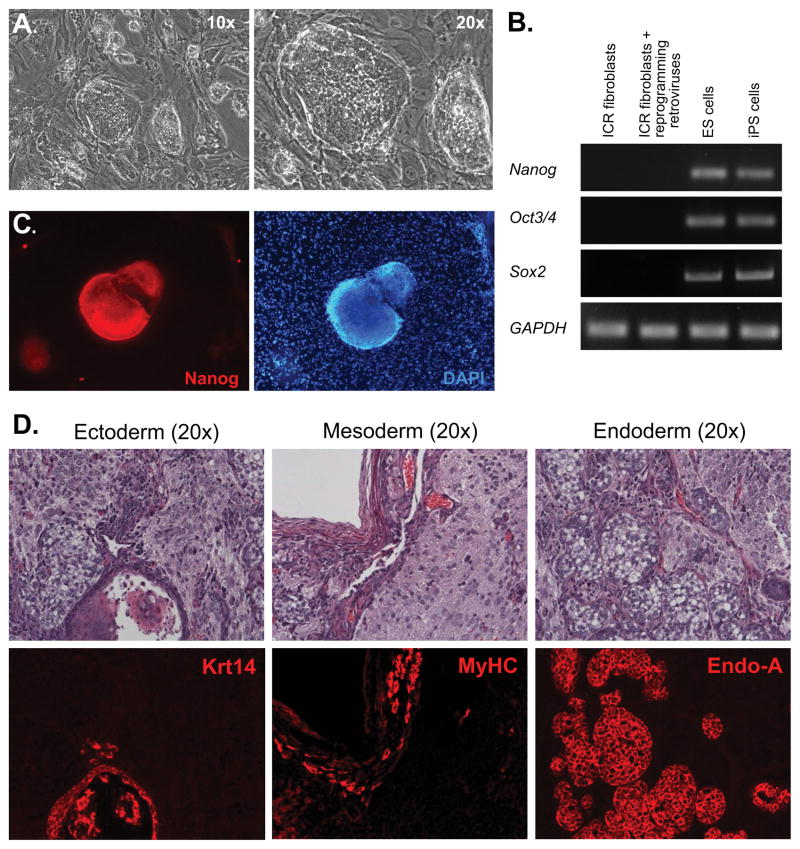
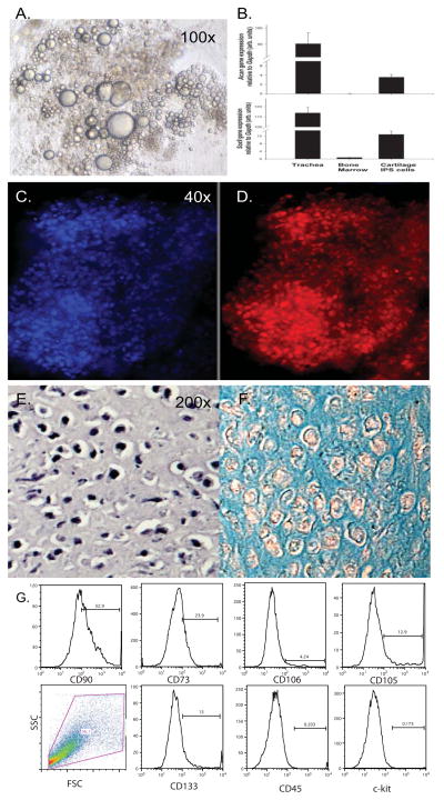
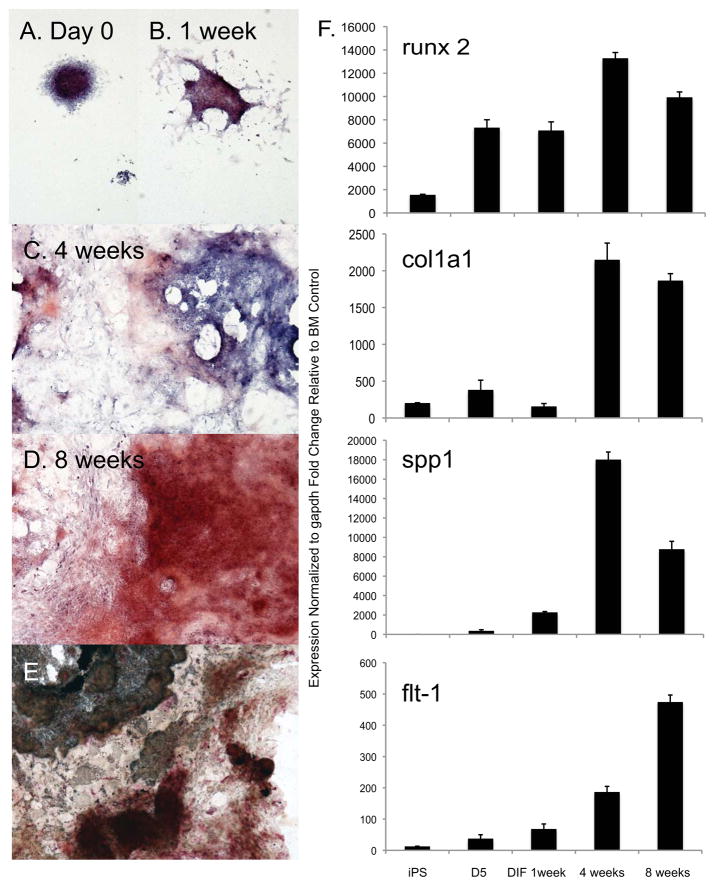
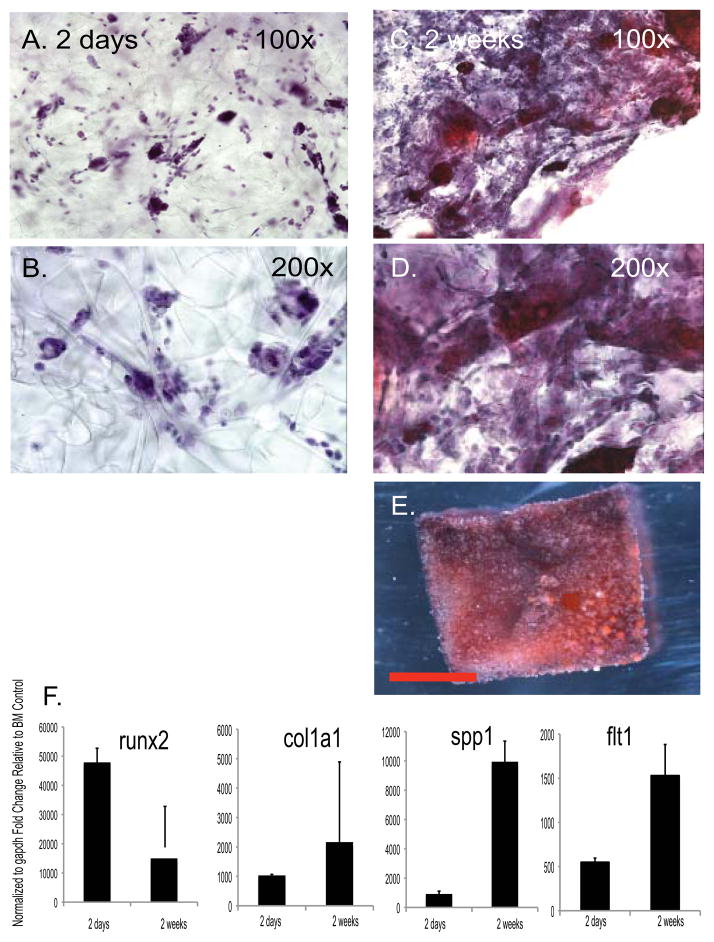
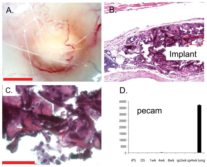
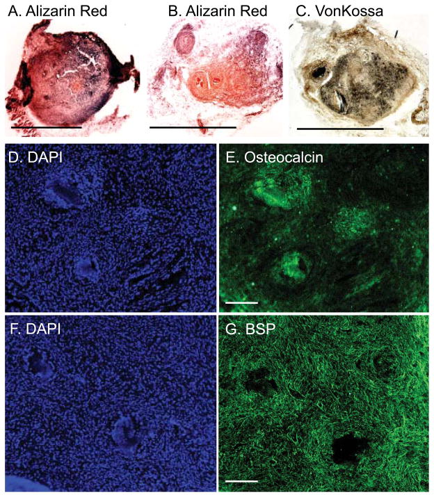
Reprogramming somatic cells into an ESC-like state, or induced pluripotent stem (iPS) cells, has emerged as a promising new venue for customized cell therapies. In this study, we performed directed differentiation to assess the ability of murine iPS cells to differentiate into bone, cartilage, and fat in vitro and to maintain an osteoblast phenotype on a scaffold in vitro and in vivo. Embryoid bodies derived from murine iPS cells were cultured in differentiation medium for 8–12 weeks. Differentiation was assessed by lineage-specific morphology, gene expression, histological stain, and immunostaining to detect matrix deposition. After 12 weeks of expansion, iPS-derived osteoblasts were seeded in a gelfoam matrix followed by subcutaneous implantation in syngenic imprinting control region (ICR) mice. Implants were harvested at 12 weeks, histological analyses of cell and mineral and matrix content were performed. Differentiation of iPS cells into mesenchymal lineages of bone, cartilage, and fat was confirmed by morphology and expression of lineage-specific genes. Isolated implants of iPS cell-derived osteoblasts expressed matrices characteristic of bone, including osteocalcin and bone sialoprotein. Implants were also stained with alizarin red and von Kossa, demonstrating mineralization and persistence of an osteoblast phenotype. Recruitment of vasculature and microvascularization of the implant was also detected. Taken together, these data demonstrate functional osteoblast differentiation from iPS cells both in vitro and in vivo and reveal a source of cells, which merit evaluation for their potential uses in orthopedic medicine and understanding of molecular mechanisms of orthopedic disease.


Conflict of interest statement

DISCLOSURE OF POTENTIAL CONFLICTS OF INTEREST

The authors indicate no potential conflicts of interest.

Figures

Figure 1
Figure 1. Generation of Mouse iPS Cells
(A): Skin fibroblasts from newborn ICR mice were transduced with retroviruses expressing reprogramming factors and cultured under ES cell conditions. After two weeks in culture, colonies exhibiting an ES cell morphology under phase-contrast microscopy emerged. (B): RT-PCR analysis with primers specific to endogenous mouse Nanog, Oct3/4 and Sox2, as well as GAPDH as a control, was performed with total RNA extracted from: Lane 1: primary ICR fibroblasts; Lane 2: ICR fibroblasts transduced with four retroviral vectors and cultured for five days; Lane 3: mouse ES cells; Lane 4: mouse iPS cells. (C): iPS cell colonies were positive for Nanog, as determined by immunofluorescence analysis (red, left panel). The feeder cells used to maintain iPS cells served as a negative control for Nanog immunofluorescence (DAPl, right panel). (D): Teratomas were formed when iPS cells were injected subcutaneously into nude mice. Top panels: H & E staining. Bottom panels were consecutive sections labeled with various antibodies representing three germ layers: Ectoderm (Krt14); Mesoderm (Myosin heavy chain from skeletal muscles (MyHC)); Endoderm (cytokeratin Endo-A). All images were taken with 20x objectives.
Figure 2
Figure 2. Differentiation of the Mesenchymal Adipocyte and Chondrocyte Lineages from iPS cells
Murine EB derived from iPS cells were treated with ATRA for 3 days to induce mesoderm followed by the treatment with adiopgenic or chondrogenic differentiation medium for an additional 4 weeks. Lipid droplets were visualized in adipocytes using phase-contrast microscopy 10x objective (A). Chondrogenesis was performed in micromass cultures and documented by qRT-PCR detection of Sox9 and Aggrecan (B) and by Aggrecan immunostaining 4x objective (D) with DAPI nuclear stain (C). The phenotype of chondrocytes was documented in paraffin sections by H&E (E) and alcian blue histochemical stains and photographs taken with the 20x objective (F). (G). Analysis of mesenchymal marker expression by dissociated iPS derived embryoid bodies following 3 day treatment with ATRA.
Figure 3
Figure 3. Functional osteoblasts can be induced from iPS cells in vitro by the presence of dexamethasone and ascorbic acid
(A–D): Murine EB derived from iPS cells were treated with ATRA for 3 days followed by osteoblast differentiation medium. Commitment to the osteoblast lineage was documented by four and eight weeks, as demonstrated by alizarin red stain, identified the increase in calcific deposit by osteoblasts (red), and nuclei were visualized with a hematoxylin counterstain (blue). (E): Von Kossa stain (black) and alkaline phosphatase colorimetric detection (red) were performed to characterize a differentiated osteoblast lineage. All images were taken with a 4x objective. (F,G): A temporal increase in expression of runx2, col1a1 and spp1 genes was confirmed by qRT-PCR analysis. BM= bone marrow.
Figure 4
Figure 4. iPS cell derived osteoblasts maintain their phenotype and deposit bone extracellular matrix on a scaffold in vitro
One-centimeter cubes of gelfoam sponge were seeded with iPS cell derived osteoblasts following 8 weeks of differentiation culture. (A, B): After 48 hours, the gelfoam scaffolds were stained with hematoxylin to confirm the presence of cells. Images were taken with 10x and 20x objective. (C,D): After 2 weeks, the scaffolds were stained with alizarin red and hematoxylin counterstain, which indicated an increase in the cells present within the scaffold, as well as matrix deposition by the osteoblasts. Images were taken with a 10x and 20x objective. (E): Nodules positive for alizarin red were detectable on gelfoam cubes in vitro after seeding with iPS cell derived osteoblasts cultured in differentiation medium for two weeks. A representative phase-contrast micrograph is presented. Scale bar = 5mm. (F): Maintenance or increased expression of runx2, col1a1 and spp1 genes was documented using qRT-PCR analysis. BM= bone marrow.
Figure 5
Figure 5. Subcutaneous implantation of osteoblast seeded scaffolds recruited vasculature in vivo
After 12 weeks, implants were identifiable (A) as surrounded by vasculature. Paraffin sections stained with hematoxylin and eosin demonstrated the subcutaneous localization of the implant (B) and the vascular supply within the implant, as evidenced by the presence of red blood cells in the lumens of microvessels (C, arrows) using phase-contrast microscopy. Scale bar = 50μM.
Figure 6
Figure 6. Osteoblast seeded scaffolds maintain their phenotype and deposit bone extracellular matrix in vivo
After 12 weeks, gelfoam scaffolds were embedded in OCT, frozen and 25mM sections analyzed. Histochemical alizarin red (A&B) and von Kossa (C) staining confirmed the presence of matrix and calcium within the nodules and hematoxylin the presence of osteoblasts. (D–G): Consecutive sections were immunoreactive to osteocalcin and bone sialoprotein. DAPI was used to label nuclei. Scale bar= 2.5mm.

References

    1. Takahashi K, et al. Induction of pluripotent stem cells from adult human fibroblasts by defined factors. Cell. 2007;131(5):861–872. - PubMed
    1. Takahashi K, Yamanaka S. Induction of pluripotent stem cells from mouse embryonic and adult fibroblast cultures by defined factors. Cell. 2006;126(4):663–676. - PubMed
    1. Wernig M, et al. In vitro reprogramming of fibroblasts into a pluripotent ES-cell-like state. Nature. 2007;448(7151):318–324. - PubMed
    1. Yamanaka S. Strategies and new developments in the generation of patient-specific pluripotent stem cells. Cell Stem Cell. 2007;1(1):39–49. - PubMed
    1. Yu J, et al. Induced pluripotent stem cell lines derived from human somatic cells. Science. 2007;318(5858):1917–1920. - PubMed

Publication types