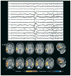
Combining EEG and fMRI in the study of epileptic discharges
- PMID: 21732941
- PMCID: PMC3753285
- DOI: 10.1111/j.1528-1167.2011.03151.x
Combining EEG and fMRI in the study of epileptic discharges
Abstract
The combining of electroencephalography (EEG) and functional magnetic resonance imaging (fMRI) is a unique noninvasive method for investigating the brain regions involved at the time of epileptic discharges. The neuronal discharges taking place during an interictal spike or spike-wave burst result in an increase in metabolism and blood flow, which is reflected in the blood oxygen-level dependent (BOLD) signal measured by fMRI. This increase is most intense in the region generating the discharge but is also present in regions affected by the discharge. On occasion, epileptic discharges result in decreased metabolism, the origin of which is only partially understood. EEG-fMRI applied to focal epilepsy results in maxima of the BOLD signal most often concordant with other methods of localization and has been shown to help in localizing epileptic foci in nonlesional frontal lobe epilepsy. It has also demonstrated the involvement of the thalamus in generalized epileptic discharges. In patients with new-onset epilepsy it could be used to evaluate the source and extent of the brain structures involved during discharges and their evolution as the disease progresses.
Wiley Periodicals, Inc. © 2011 International League Against Epilepsy.
Figures


References
-
- Aghakhani Y, Bagshaw AP, Benar CG, Hawco C, Andermann F, Dubeau F, Gotman J. fMRI activation during spike and wave discharges in idiopathic generalized epilepsy. Brain. 2004;127:1127–1144. - PubMed
-
- Benar CG, Gross DW, Wang Y, Petre V, Pike B, Dubeau F, Gotman J. The BOLD response to interictal epileptiform discharges. Neuroimage. 2002;17:1182–1192. - PubMed
-
- Benar CG, Grova C, Kobayashi E, Bagshaw AP, Aghakhani Y, Dubeau F, Gotman J. EEG-fMRI of epileptic spikes: concordance with EEG source localization and intracranial EEG. Neuroimage. 2006;30:1161–1170. - PubMed

