

Functional exchangeability of the nuclear localization signal (NLS) of capsid protein between PCV1 and PCV2 in vitro: Implications for the role of NLS in viral replication
- PMID: 21733152
- PMCID: PMC3145596
- DOI: 10.1186/1743-422X-8-341
Functional exchangeability of the nuclear localization signal (NLS) of capsid protein between PCV1 and PCV2 in vitro: Implications for the role of NLS in viral replication
Abstract
Background: Porcine circovirus type 2 (PCV2) is believed to be the primary causative agent of postweaning multisystemic wasting syndrome (PMWS). It is supposed that capsid protein of PCV may contribute to replication control via interaction between Cap and Rep in the nucleoplasm. In this study, we described the construction and in vitro characterization of NLS-exchanged PCV DNA clones based on a PMWS-associated PCV2b isolate from China to determine the role of ORF2 NLS in PCV replication.
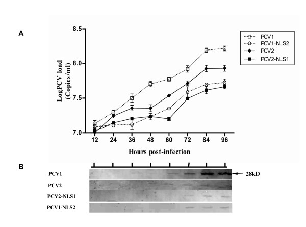
Results: The PCV1, PCV2, PCV2-NLS1 and PCV1-NLS2 DNA clone were generated by ligating a copy of respective genome in tandem with a partial duplication. The PCV2-NLS1 and PCV1-NLS2 DNA clone contained a chimeric genome in which the ORF2 NLS was exchanged. The four DNA clones were all confirmed to be infectious in vitro when transfected into PK-15 cells, as PCV capsid protein were expressed in approximately 10-20% of the transfected cells. The in vitro growth characteristics of the DNA clones were then determined and compared. All the recovered progeny viruses gave rise to increasing infectious titers during passages and were genetically stable by genomic sequencing. The chimeric PCV1-NLS2 and PCV2-NLS1 viruses had the final titers of about 104.2 and 103.8 TCID50/ml, which were significantly lower than that of PCV1 and PCV2 (105.6 and 105.0 TCID50/ml, respectively). When the ORF2 NLS exchanged, the mutant PCV2 (PCV2-NLS1) still replicated less efficiently and showed lower infectious titer than did PCV1 mutant (PCV1-NLS2), which was consistent with the distinction between wild type PCV1 and PCV2.
Conclusions: Recovery of the chimeiric PCV1-NLS2 and PCV2-NLS1 progeny viruses indicate that the nuclear localization signal sequence of capsid protein are functionally exchangeable between PCV1 and PCV2 with respect to the role of nuclear importing and propagation. The findings also reveal that ORF2 NLS play an accessory role in the replication of PCV. However, we found that ORF2 NLS was not responsible for the distinction of in vitro growth characteristic between PCV1 and PCV2. Further studies are required to determine the in vivo viral replication and pathogenicity of the NLS chimeric DNA clones.
Figures
Similar articles
-
In vivo characterization of chimeric PCV DNA clones containing heterogeneous capsid protein nuclear localization signals (NLS).Virol J. 2013 Jan 7;10:16. doi: 10.1186/1743-422X-10-16. Virol J. 2013. PMID: 23294939 Free PMC article.
-
Immunogenicity and pathogenicity of chimeric infectious DNA clones of pathogenic porcine circovirus type 2 (PCV2) and nonpathogenic PCV1 in weanling pigs.J Virol. 2003 Oct;77(20):11232-43. doi: 10.1128/jvi.77.20.11232-11243.2003. J Virol. 2003. PMID: 14512571 Free PMC article.
-
Epitope mapping of the major capsid protein of type 2 porcine circovirus (PCV2) by using chimeric PCV1 and PCV2.J Virol. 2004 Aug;78(15):8135-45. doi: 10.1128/JVI.78.15.8135-8145.2004. J Virol. 2004. PMID: 15254185 Free PMC article.
-
Porcine circoviruses--small but powerful.Virus Res. 2009 Aug;143(2):177-83. doi: 10.1016/j.virusres.2009.02.009. Epub 2009 Feb 26. Virus Res. 2009. PMID: 19647885 Review.
-
Genotypic diversity and immunological implications of porcine circovirus: Inspiration from PCV1 to PCV4.Microb Pathog. 2024 Nov;196:106997. doi: 10.1016/j.micpath.2024.106997. Epub 2024 Oct 5. Microb Pathog. 2024. PMID: 39369754 Review.
Cited by
-
Structural insight into the type-specific epitope of porcine circovirus type 3.Biosci Rep. 2020 Jun 26;40(6):BSR20201109. doi: 10.1042/BSR20201109. Biosci Rep. 2020. PMID: 32458997 Free PMC article.
-
Genome architecture changes and major gene variations of Andrias davidianus ranavirus (ADRV).Vet Res. 2013 Oct 21;44(1):101. doi: 10.1186/1297-9716-44-101. Vet Res. 2013. PMID: 24143877 Free PMC article.
-
Advances in Crosstalk between Porcine Circoviruses and Host.Viruses. 2022 Jun 28;14(7):1419. doi: 10.3390/v14071419. Viruses. 2022. PMID: 35891399 Free PMC article. Review.
-
In vivo characterization of chimeric PCV DNA clones containing heterogeneous capsid protein nuclear localization signals (NLS).Virol J. 2013 Jan 7;10:16. doi: 10.1186/1743-422X-10-16. Virol J. 2013. PMID: 23294939 Free PMC article.
-
Highly efficient cellular uptake of a cell-penetrating peptide (CPP) derived from the capsid protein of porcine circovirus type 2.J Biol Chem. 2018 Sep 28;293(39):15221-15232. doi: 10.1074/jbc.RA118.004823. Epub 2018 Aug 14. J Biol Chem. 2018. PMID: 30108178 Free PMC article.
References
-
- Meehan BM, McNeilly F, Todd D, Kennedy S, Jewhurst VA, Ellis JA, Hassard LE, Clark EG, Haines DM, Allan GM. Characterization of novel circovirus DNAs associated with wasting syndromes in pigs. J Gen Virol. 1998;79:2171–2179. - PubMed
-
- Segales J, Domingo M. Postweaning multisystemic wasting syndrome (PMWS) in pigs: a review. Vet Q. 2002;24:109–124. - PubMed
Publication types
MeSH terms
Substances
LinkOut - more resources
Full Text Sources
Miscellaneous
