Review
. 2011 Nov;91(6):497-509.
doi: 10.1016/j.tube.2011.03.007. Epub 2011 Jul 6.

Pathology of post primary tuberculosis of the lung: an illustrated critical review

Affiliations
Review

Pathology of post primary tuberculosis of the lung: an illustrated critical review

Robert L Hunter. Tuberculosis (Edinb). 2011 Nov.

Abstract

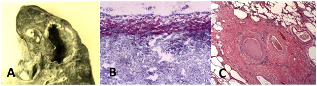
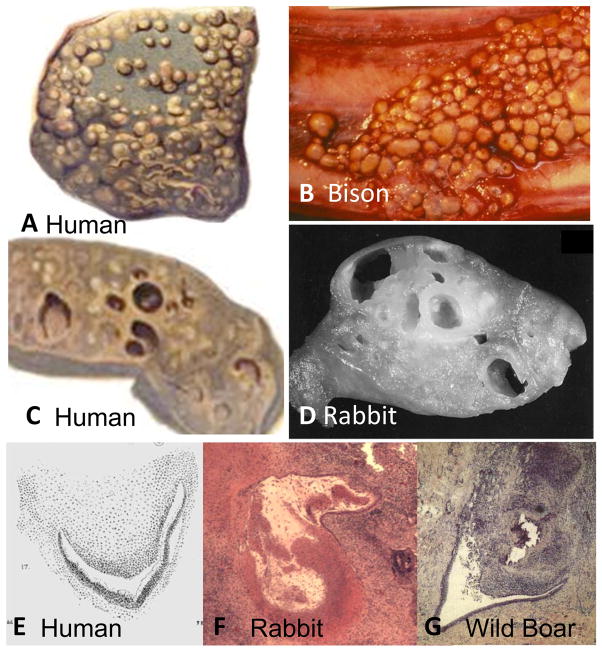
Post primary tuberculosis occurs in immunocompetent adults, is restricted to the lungs and accounts for 80% of all clinical cases and nearly 100% of transmission of infection. The supply of human tissues with post primary tuberculosis plummeted with the introduction of antibiotics decades before the flowering of research using molecular methods in animal models. Unfortunately, the paucity of human tissues prevented validation of the models. As a result, it is a paradigm of contemporary research that caseating granulomas are the characteristic lesion of all tuberculosis and that cavities form when they erode into bronchi. This differs from descriptions of the preantibiotic era when many investigators had access to thousands of cases. They reported that post primary tuberculosis begins as an exudative reaction: a tuberculous lipid pneumonia of foamy alveolar macrophages that undergoes caseation necrosis and fragmentation to produce cavities. Granulomas in post primary disease arise only in response to old caseous pneumonia and produce fibrosis, not cavities. We confirmed and extended these observations with study of 104 cases of untreated tuberculosis. In addition, studies of the lungs of infants and immunosuppressed adults revealed a second type of tuberculous pneumonia that seldom produces cavities. Since the concept that cavities arise from caseating granulomas was supported by studies of animals infected with Mycobacterium bovis, we investigated its pathology. We found that M. bovis does not produce post primary tuberculosis in any species. It only produces an aggressive primary tuberculosis that can develop small cavities by erosion of caseating granulomas. Consequently, a key unresolved question in the pathogenesis of tuberculosis is identification of the mechanisms by which Mycobacterium tuberculosis establish a localized safe haven in alveolar macrophages in an otherwise solidly immune host where it can develop conditions for formation of cavities and transmission to new hosts.


Conflict of interest statement

Competing interests: No confl

Figures

Figure 1
Figure 1. Caseating granulomas of primary tuberculosis
A central core of necrotic lipid rich material is surrounded by foamy macrophages, epitheloid macrophages and lymphocytes. Three examples of caseating granuloma are shown: (A) shows an early caseating granuloma in the lung of a person with miliary tuberculosis (H & E Stain 100× Magnification). (B) A cluster of caseating granulomas that was mistaken for a cancer and surgically removed. Cuffs of blue staining lymphocytes surrounding caseous necrotic material are particularly evident (H & E Stain 1× Magnification). (C) An older lesion in a human lung showing fibrous capsule and necrotic center. Epitheloid cells and a giant cell (arrow) indicate that it is still active (H & E Stain 100× Magnification).
Figure 2
Figure 2. Histopathology of developing post-primary tuberculosis
(A), Initially, the alveoli are filled with macrophages while the alveolar walls have increased numbers of lymphocytes. The macrophages are foamy due to accumulation of lipid. Such lesions may develop in several ways. In some areas (B), the cellular debris and fibrin predominate. In others, (C) the macrophages become increasingly lipid rich. Langhans giant cells may be found (D). Eventually, many cells within the alveoli undergo caseation necrosis (E). The process may involve larger areas of the lung or single alveoli. Most such lesions may spontaneously resolve with replacement of alveolar cells with fibrin and fibrous tissue (F). Resolving lesions eventually form fibrous scars in the apices of lungs (H & E Stains 100 to 400× Magnification).
Figure 3
Figure 3. Bronchial occlusion in post-primary tuberculosis
‘Tree and bud’ pattern is the characteristic CAT scan appearance of early post-primary tuberculosis , (A). The tree twigs in the radiograph are bronchi filled with infected cells and debris while the buds are areas of tuberculous pneumonia in alveoli. The section (B, C) shows a bronchi occluded by such material. Bronchial obstruction has been reported by many authors in 100% of cases of post-primary pulmonary tuberculosis , , , (H & E Stain 100× Magnification)
Figure 4
Figure 4. Distribution of AFB and mycobacterial antigen in post primary TB
Very few AFB are found in foamy alveolar macrophages of developing post primary tuberculosis. Their numbers increase markedly with the development of necrosis. (A) Section of lung showing masses of AFB in an alveolus with caseous necrosis, but not in adjacent viable alveoli (acid fast stain 40X). (B). High magnification of AFB associated with necrotic cells and debris (acid fast stain 400X). (C) A single AFB in a viable macrophage (acid fast stain 400X). (D) Demonstration of abundant mycobacterial antigens in viable cells that have no AFB (Immunohistochemical stain for MTB 400×).
Figure 5
Figure 5. Formation of Cavities
In its classic occurrence, tuberculous cavities form rapidly following onset of acute febrile pneumonia. (A) A developing cavity containing necrotic lung tissue. The wall of the cavity is composed of a layer of necrosis overlying lipid pneumonia. The cavity contains fragments of necrotic lung (Arrows) (H & E Stain 4× Magnification). This lung was positive for MTB by PCR and AFB even though the number of organisms was small. (B) Vasculitis in a small artery in the lung. The vessel wall is thickened, vacuolated and infiltrated with lymphocytes. The lumen is largely occluded (H & E Stain 100× Magnification). (C) Necrotic lung tissue within a bronchus of a person with acutely developing cavitary tuberculosis (H & E Stain 40× Magnification).
Figure 6
Figure 6. Maturation of Cavities
Cavities frequently develop as thin walled structures with little inflammation and masses of organisms growing as a pellicle on the inner surface. (A) A cavity in the upper lobe of the lung with connection to a bronchus. The disease was largely asymptomatic and the patient died of unrelated illness. The wall of the cavity was composed of fibrous tissue and a layer of necrotic debris. (B) The surface demonstrated massive numbers of acid-fast organisms (acid-fast stain 200X). No organisms or evidence of active tuberculosis were present anywhere else in the body. (C) Other areas of the lung demonstrated scars of healed tuberculosis with no evidence of recent activity (H & E Stain 100× Magnification).
Figure 7
Figure 7. Chronic fibrocaseous tuberculosis
Masses of caseous pneumonia that do not soften and fragment to produce cavities may simply persist in a relatively inactive state. In this case, a necrotic upper lobe of the lung with caseous pneumonia has become mummified (A). Microscopically it consists of caseous pneumonia adjacent to viable areas of lipid-rich tuberculous pneumonia (B,C) (H & E Stain 100–400× Magnification). More often the aging caseous pneumonia becomes surrounded by granulomatous tissue (D). The central necrotic core retains the structure of alveoli with caseous pneumonia that is surrounded by an active granulomatous process with epitheloid cells, giant cells and lymphocytes, but no foamy macrophages. Fibrosis surrounds smaller areas of caseous pneumonia (E) or extends across large areas of lung with larger lesions to produce pulmonary fibrosis (F). Multiple layers of fibrosis demonstrate that the lesion has progressed and regressed on multiple occasions (G). (Trichrome Stains 40× Magnification). Rarely, erosion of caseating granulomas into cavities is seen (G). However, the lesions are always small and it is seldom clear whether the cavity eroded into the granuloma or visa versa.
Figure 8
Figure 8. Tuberculosis in immunosuppressed people
Case 1. (A–C) Tuberculosis associated with HIV. Multiple areas of the lung demonstrated tuberculous pneumonia in which alveoli contained macrophages with many AFB (insert) but very little lipid (A). Alveolar wall contained many lymphocytes. Large areas of the tuberculous pneumonia had undergone recent necrosis (B). Still others areas of the lung demonstrated caseating granulomas of miliary tuberculosis (C) (H & E Stain 100–400 × Magnification, Insert Acid-fast stain 1000× magnification). Case 2 (D–F): Cavitary tuberculosis in an infant. A 9-month-old child died of rapidly progressive tuberculosis. The lung contained many caseating granulomas, large foci of tuberculous pneumonia and cavities (D arrow). The lung adjacent to the cavities contained necrotizing tuberculous pneumonia (E). The viable areas demonstrated tuberculous pneumonia with cells that contained very little lipid (F). Cavities were found exclusively in the areas of tuberculosis pneumonia, not in the nodule lesions of caseating granulomas. (H & E Stain 40–200× Magnification). Case 3. (G–H) Tuberculosis in an immunosuppressed individual. A man with rheumatoid arthritis receiving large doses of steroids died of rapidly progressive tuberculosis. Lesions of the lung were a caseous pneumonia with few macrophages or lymphoid cells but much edema and fibrinous exudate within alveoli (G). Massive numbers of acid-fast organisms were present in the areas of acute necrosis (H) (H & E Stain 40 – 200× Magnification, Acid-fast stain 400 × magnification).
Figure 9
Figure 9. Pathology of M. bovis tuberculosis in humans and animals
The characteristic lesions of M. bovis tuberculosis are nodules up to 2 cm in diameter that grow on serosal surfaces including the diaphragm, lung, and peritoneum, in addition to deep within organs. The top two figures show lesions on the pleural surfaces of a human (A) and bison (B). The lower figures show cut surfaces of cavitary lung lesions in a human (C) and rabbit (D). The cavities are caseating granulomas that have undergone central softening and erosion into adjacent structures to produce cavities. Cavities caused by M. bovis in a human (E) rabbit (F) and wild boar (G) and have similar characteristics. Caseating granulomas have eroded into bronchi. The images of rabbit and wild boar lesions show central softening and liquefaction. The only available image of a human lesion shows similar morphology . (Wild boar image from Gortazar with permission . The rabbit slide from Converse by permission ) (H & E Stain 40× Magnification). (Human Pictures are from Creighton, 1881 Bison picture from U Manitoba by permission).

References

    1. Dubos R, Dubos G. Tuberculosis, Man, and Society. Brunswick, NJ: Rutgers Univesity Press; 1987. The White Plague.
    1. Garay S. Pulmonary Tuberculosis. In: Garay WRS, editor. Tuberculosis. Chapter 32. Boston: Little Brown & Co; 1996. pp. 373–412.
    1. Leong FJ, Dartois V, Dick T. A Color Atlas of Comparative Pathology of Pulmonary Tuberculosis. New York: CRC Press; 2011.
    1. McGrath JW. Social networks of disease spread in the lower Illinois valley: a simulation approach. Am J Phys Anthropol. 1988;77:483–96. - PubMed
    1. Donald PR, Marais BJ, Barry CE., 3rd Age and the epidemiology and pathogenesis of tuberculosis. Lancet. 2010;375:1852–4. - PubMed

Publication types