. 2011 Sep 9;286(36):31650-60.
doi: 10.1074/jbc.M111.253401. Epub 2011 Jul 6.

Molecular insights into DNA polymerase deterrents for ribonucleotide insertion

Affiliations

Molecular insights into DNA polymerase deterrents for ribonucleotide insertion

Nisha A Cavanaugh et al. J Biol Chem. .

Abstract

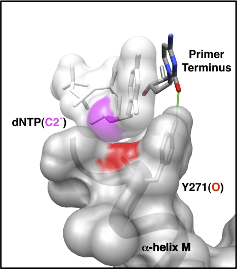
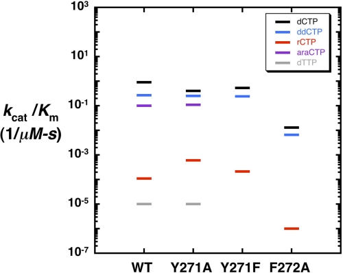
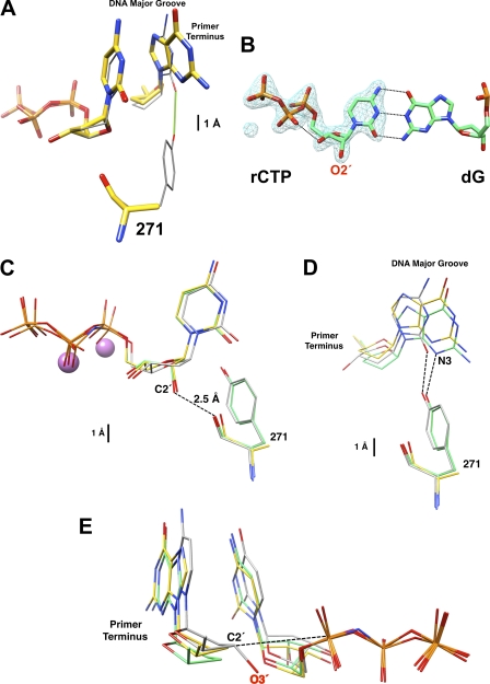
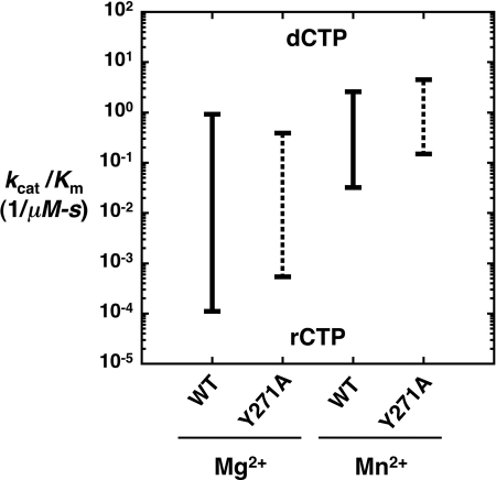
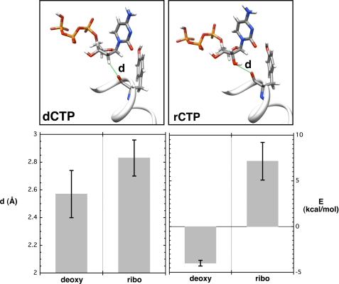
DNA polymerases can misinsert ribonucleotides that lead to genomic instability. DNA polymerase β discourages ribonucleotide insertion with the backbone carbonyl of Tyr-271; alanine substitution of Tyr-271, but not Phe-272, resulted in a >10-fold loss in discrimination. The Y271A mutant also inserted ribonucleotides more efficiently than wild type on a variety of ribonucleoside (rNMP)-containing DNA substrates. Substituting Mn(2+) for Mg(2+) decreased sugar discrimination for both wild-type and mutant enzymes primarily by increasing the affinity for rCTP. This facilitated crystallization of ternary substrate complexes of both the wild-type and Y271A mutant enzymes. Crystallographic structures of Y271A- and wild type-substrate complexes indicated that rCTP is well accommodated in the active site but that O2' of rCTP and the carbonyl oxygen of Tyr-271 or Ala-271 are unusually close (∼2.5 and 2.6 Å, respectively). Structure-based modeling indicates that the local energetic cost of positioning these closely spaced oxygens is ∼2.2 kcal/mol for the wild-type enzyme. Because the side chain of Tyr-271 also hydrogen bonds with the primer terminus, loss of this interaction affects its catalytic positioning. Our results support a model where DNA polymerase β utilizes two strategies, steric and geometric, with a single protein residue to deter ribonucleotide insertion.


Figures

FIGURE 1.
FIGURE 1.
Molecular interactions of Tyr-271 with the incoming nucleotide and primer terminus. Tyr-271 of α-helix M forms part of the dNTP-binding pocket and interacts with the minor groove edge of the DNA substrate. In the closed polymerase conformation, the side chain of Tyr-271 hydrogen bonds with the minor groove edge of the primer terminal base (green line). The backbone carbonyl of Tyr-271 (red) is within 3.5 Å of C2′ (magenta) of the incoming nucleotide. The van der Waals semi-transparent surfaces (gray) of the incoming nucleotide and α-helix M are shown. In the open polymerase conformation, Tyr-271 interacts with the minor groove edge of the templating base (not shown).
FIGURE 2.
FIGURE 2.
Substrate discrimination by human DNA polymerase β mutants. A discrimination plot where the catalytic efficiencies (−) for nucleotide insertion (Table 1) are plotted on a log ordinate scale. Nucleotide insertion was measured on a single-nucleotide gapped substrate with a templating “dG” as described under “Experimental Procedures.” Discrimination is proportional to the distance between a colored bar and a black bar (corresponding to correct insertion of dCTP) for each enzyme. Thus, the greater the distance between catalytic efficiencies, the greater the discrimination. The catalytic efficiencies for ddCTP (blue), araCTP (purple), rCTP (red), and dTTP (gray) are plotted for wild-type pol β and Y271A, Y271F, and F272A mutants.
FIGURE 3.
FIGURE 3.
Structural characterization of dCTP/rCTP in the confines of wild-type and Y271A DNA polymerase β. A, active site comparison between superimposed ternary complex structures with a correct active site base pair for wild-type (PDB ID 2FMP; gray carbons) (26) and Y271A mutant (yellow carbons) of pol β. The incoming nucleotide (ddCTP, wild type; dCTP, Y271A) is shown along with the dideoxy-terminated primer terminus and residue 271. Loss of the hydrogen bond (green) with the minor groove edge of the base of the primer terminus in the Y271A structure results in a modest displacement into the DNA major groove. B, FoFc simulated annealing electron density omit map (blue) contoured at 5.0σ showing electron density corresponding to rCTP bound to wild-type pol β. The O2′ of the ribose sugar is clearly observed and the base forms Watson-Crick base pairs with the templating deoxyguanine (dG). C, comparison of the incoming nucleotides with superimposed structures of wild-type enzyme with an incoming nucleotide analog of dTTP (PDB code 2FMS, gray) (26) and wild-type (green) and Y271A mutant (yellow) pol β with an incoming rCTP. The structures were superimposed using all Cαs. The root mean square deviations are given in the text. Residue 271 (tyrosine or alanine) is also illustrated. The O2′ of the ribose sugar of rCTP is unusually close (2.54 Å) to the carbonyl backbone of residue 271 in the wild-type structure. The two purple spheres represent the active site Mn2+ ions from the wild-type structure with an incoming rCTP. D, from the position of the primer terminus in the superimposed structures from C, the loss of the hydrogen bond between the side chain of Tyr-271 and the minor groove edge of the base of the primer terminus results in a similar displacement into the major groove as observed with a correct incoming nucleotide. This is most easily seen by looking at the position of N3 of the guanine base in the structures of wild-type and mutant enzymes with an incoming rCTP. E, to compare the relative position of C3′/O3′ of the primer terminus relative to Pα of the incoming nucleotide, the triphosphate portion of wild-type and mutant enzymes was superimposed. The structures with an incoming rCTP do not include O3′ of the primer terminus (i.e. dideoxy-terminated), whereas the wild-type enzyme with an inert dTTP analog includes O3′ of the primer terminus and represents the reference structure for ideal active site geometry. The relative position of the primer termini, as judged by the position of C2′, suggests that the Y271A mutant might achieve a modestly better active site geometry with an incoming rCTP than wild-type enzyme.
FIGURE 4.
FIGURE 4.
Effect on Mn2+ on ribonucleotide discrimination. A discrimination plot comparing catalytic efficiencies (−) for wild-type and Y271A pol β in the presence of Mg2+ and Mn2+. Nucleotide (dCTP or rCTP) insertion was measured on a single-nucleotide gapped substrate with a templating dG as described under “Experimental Procedures.” Discrimination is proportional to the distance between the two catalytic efficiencies for wild type (solid line) and Y271A (dotted line).
FIGURE 5.
FIGURE 5.
Interaction of the carbonyl of Tyr-271 with an incoming deoxy- or ribonucleotide. MD simulations were performed as described under “Experimental Procedures,” and the final unconstrained runs (10 ns) were carried out with time steps of 1 fs. The upper illustrations highlight the distance (d, green line) of the interacting atoms that are monitored for dCTP (left panel) and rCTP (right panel). A portion of α-helix M with Tyr-271 is also illustrated. The average dCTP(H2′)/Tyr-271(O) and rCTP(O2′)/Tyr-271(O) distances and local interaction energies (E) are plotted below.
FIGURE 6.
FIGURE 6.
Comparing ribonucleotide discrimination for wild-type and mutant DNA polymerases from different families. Ribonucleotide discrimination has been typically attributed to a steric gate residue in the active site (10). Discrimination plots compare catalytic efficiencies (−) for dCTP and rCTP insertion by wild-type (solid line) and mutant DNA polymerases (dotted line) from A-, B-, X-, and Y-family DNA polymerases (, , –46). Ribonucleotide discrimination is proportional to the distance between the catalytic efficiencies for dCTP and rCTP. A, ribonucleotide discrimination of wild-type and mutant DNA polymerases when the steric gate or fence residue is removed. B, ribonucleotide discrimination of wild-type and mutant DNA polymerases when a nonsteric gate residue is altered.

References

    1. Tsai Y. C., Johnson K. A. (2006) Biochemistry 45, 9675–9687 - PMC - PubMed
    1. Batra V. K., Beard W. A., Hou E. W., Pedersen L. C., Prasad R., Wilson S. H. (2010) Nat. Struct. Mol. Biol. 17, 889–890 - PMC - PubMed
    1. Ferraro P., Franzolin E., Pontarin G., Reichard P., Bianchi V. (2010) Nucleic Acids Res. 38, e85. - PMC - PubMed
    1. Traut T. W. (1994) Mol. Cell. Biochem. 140, 1–22 - PubMed
    1. Cavanaugh N. A., Beard W. A., Wilson S. H. (2010) J. Biol. Chem. 285, 24457–24465 - PMC - PubMed

Publication types

LinkOut - more resources