. 2011 Sep 2;286(35):30274-30283.
doi: 10.1074/jbc.M111.240309. Epub 2011 Jul 6.

Stalled proteasomes are directly relieved by P97 recruitment

Affiliations

Stalled proteasomes are directly relieved by P97 recruitment

Elada Isakov et al. J Biol Chem. .

Abstract

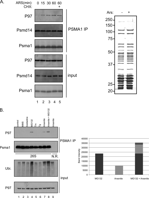
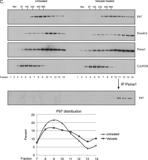
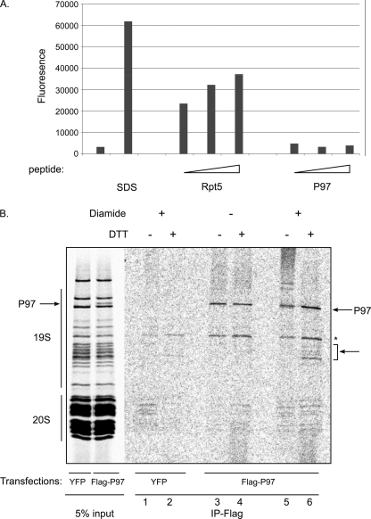
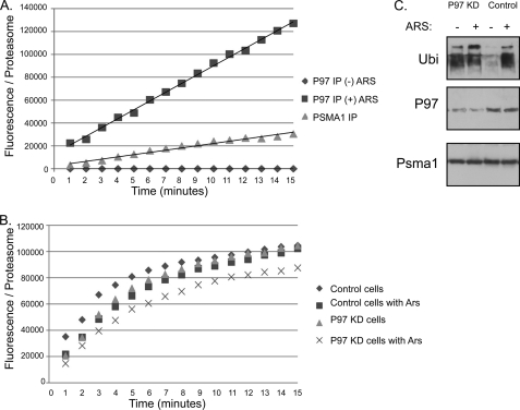
The 26 S proteasome is the eukaryotic protease responsible for the degradation of most cellular proteins. As such it accommodates the ability to function under diverse conditions that the cell may encounter. This function is supported by various adaptors that modulate various aspects in protein degradation, these include regulation of substrate delivery, deubiquitination, unfolding, and 20 S gate dilation. Here we show a new functional complex between the P97 and the proteasome that is assembled in response to proteasomal impairment. This entails P97 binding to the 26 S proteasome via the 19 S particle thereby forming an additional hexameric ATPase ring to relieve repression. P97-bound proteasomes showed selective binding toward the Npl4-ufd1 P97 co-factors, indicating a unique cellular role for P97 binding to proteasomes. P97-bound proteasomes display enhanced activity, showing a relief in proteolysis impairment. Our findings place P97 directly in non-ERAD proteasomal functions and establish a new checkpoint in UPS impairment. The ability to modulate proteasome activity and properly respond to protein misfolding, is of great importance in cellular regulation.


Figures

FIGURE 1.
FIGURE 1.
Proteasome-P97 interaction. A, left panel shows a time course induction of P97-proteasome interaction upon arsenite treatment (0.5 mm). Untreated or arsenite-treated lysates were subjected to PSMA1 (26 S) IP followed by a PSMD14 and P97 immunoblots. Cycloheximide (10 μg/ml) was added to prevent de novo protein synthesis. Right panel indicates the protein content (as evaluated by Coomassie-stained SDS-PAGE) of the 26 S proteasomes purified from control and arsenite-treated cells. Input represents 50 μg of total cell extract while the IP was performed using 2 mg of cell extract. B, HEK 293 cells were treated with several stress agents and subjected to PSMA1 (26 S) IP and P97 immunoblot. The stress agents indicated are: 2, the electrophile methylmethanesulfonate (MMS, 2.4 mm 1 h); 3, the amino acid analog Azetidine (5 mm, overnight); 4, proteasome inhibitor MG132 (10 μm, 1 h); 5, the ER stress-inducing agents tunicamycin (Tm, 20 μg/ml, 1 h); 6, thapsigargin (Tg, 2 μm, 1 h) 7, arsenite (0.5 mm, 1 h); and 8, MG132 (10 μm, 1 h)+arsenite (0.5 mm, 1 h). Lane 1 represents the untreated cells. As a negative control we used a non-relevant IP against cells treated with arsenite. Right panel shows the quantitation of the P97 signal indicating the accumulative effect of MG132 and arsenite. Input represents 50 μg of total cell extract while the IP was performed using 2 mg of cell extract. C, immunoblot of P97, the 19S lid component PSMD14, the 20 S subunit PSMA1 and GAPDH in fractions of glycerol gradients prepared from untreated and Velcade-treated cells (1 h 10 μm). The immunoblot of P97 in PSMA1 immunoprecipitations from fractions of glycerol gradients is shown in the bottom panel. The migration of complexes of known size is indicated above. The migration of P97 from the indicated fractions was quantified and presented as fractions from the total signal. Note the increase in P97 in the HMW fractions upon Velcade treatment. GAPDH IB served as a nonspecific migration control.
FIGURE 1.
FIGURE 1.
Proteasome-P97 interaction. A, left panel shows a time course induction of P97-proteasome interaction upon arsenite treatment (0.5 mm). Untreated or arsenite-treated lysates were subjected to PSMA1 (26 S) IP followed by a PSMD14 and P97 immunoblots. Cycloheximide (10 μg/ml) was added to prevent de novo protein synthesis. Right panel indicates the protein content (as evaluated by Coomassie-stained SDS-PAGE) of the 26 S proteasomes purified from control and arsenite-treated cells. Input represents 50 μg of total cell extract while the IP was performed using 2 mg of cell extract. B, HEK 293 cells were treated with several stress agents and subjected to PSMA1 (26 S) IP and P97 immunoblot. The stress agents indicated are: 2, the electrophile methylmethanesulfonate (MMS, 2.4 mm 1 h); 3, the amino acid analog Azetidine (5 mm, overnight); 4, proteasome inhibitor MG132 (10 μm, 1 h); 5, the ER stress-inducing agents tunicamycin (Tm, 20 μg/ml, 1 h); 6, thapsigargin (Tg, 2 μm, 1 h) 7, arsenite (0.5 mm, 1 h); and 8, MG132 (10 μm, 1 h)+arsenite (0.5 mm, 1 h). Lane 1 represents the untreated cells. As a negative control we used a non-relevant IP against cells treated with arsenite. Right panel shows the quantitation of the P97 signal indicating the accumulative effect of MG132 and arsenite. Input represents 50 μg of total cell extract while the IP was performed using 2 mg of cell extract. C, immunoblot of P97, the 19S lid component PSMD14, the 20 S subunit PSMA1 and GAPDH in fractions of glycerol gradients prepared from untreated and Velcade-treated cells (1 h 10 μm). The immunoblot of P97 in PSMA1 immunoprecipitations from fractions of glycerol gradients is shown in the bottom panel. The migration of complexes of known size is indicated above. The migration of P97 from the indicated fractions was quantified and presented as fractions from the total signal. Note the increase in P97 in the HMW fractions upon Velcade treatment. GAPDH IB served as a nonspecific migration control.
FIGURE 2.
FIGURE 2.
The P97-proteasome complex contains both the 19 S and the 20 S particles. A, immunoblots of P97, Psmd14, and YFP after YFP IP's from CHO clones expressing the indicated proteins obtained from untreated or Velcade-treated cells. The right panels reveal the glycerol gradient YFP immunoblots of the various clones. B, 293 cells untreated or MG132 treated were lysed and purified with P97 antibody, P97 IP followed by Rpt1 and Psma1 immunoblots. As a negative control, we used a non-relevant IP (NR IP).
FIGURE 3.
FIGURE 3.
P97 binds the 19 S particle of the proteasome. A, 0.01% SDS and several concentrations of peptides corresponding to the C-terminal residues of P97 and Rpt5 (50, 200, 400 μm) were incubated with 20 S proteasomes and activity against the N-succinyl-LLVY-AMC (100 μm) fluoregenic peptide was measured. B, autoradiograph of a SDS-PAGE of metabolically labeled proteins purified by glycerol gradient centrifugation followed by PSMA1 immunoprecipitation from cells transfected with Flag P97 or YFP-treated with MG132. The purified complex was displayed as is (lanes 1 and 2) or exposed to diamide (100 μm), followed by disruption in 2% SDS. Samples were diluted and immunoprecipited with an anti-Flag antibody as indicated (lanes 3–6). Note the appearance of several bands with the approximate mass of 50 kDa in a P97 and diamide-dependent manner.
FIGURE 4.
FIGURE 4.
Mapping of the proteasome binding region of P97. A, full-length p97 or truncated varients were expressed as FLAG-tagged fusion proteins in HEK 293T cells treated with MG132. Expression of FLAG-tagged p97 proteins was detected by immunoblotting before (input left panel) and after IP of PSMA1 (right panel). Note the importance of D1 and D2 domains for P97-proteasome complex formation. B, wild type p97 or truncated proteins were expressed in TNT reticulocyte expression system and subjected to glycerol gradient centrifugation. The collected fractions were resolved on SDS-PAGE and exposed to autoradiography. The hexameric form of P97 proteins is indicated. Flag P97 linker +D2 (461–806) and Flag P97 ΔD2 (1–480) fail to form hexamers.
FIGURE 5.
FIGURE 5.
P97 cofactors specificity and requirement in P97-proteasome co-purifications. A, P97 cofactors were expressed as FLAG-tagged fusion proteins in HEK 293T cells untreated or treated with Velcade (as indicated). Lysates were subjected to a direct immunoblot (input) or immunprecipitated with a PSMA1 antibody. P97 and FLAG expressed protein content co-purified with PSMA1 were evaluated on the immune-purified material (Psma1 IP). B, stable clones expressing a Ufd1 knock-down expression vector were produced to evaluate the requirement of Ufd1 toward P97-proteasome co-purification. Proteasomal-P97 co-purifications were performed as described in Fig. 1.
FIGURE 6.
FIGURE 6.
Induced activity and role of the P97-proteasome complex. A, digestion of a small, unstructured peptide (N-succinyl-LLVY-AMC), monitored by fluorescence in samples of proteasomes purified by P97 immunoprecipitation from cells untreated or arsenite-treated. PSMA1 IP served as a reference for the activity of conventional proteasomes and GST purifications as a background control. All observed activity was inhibited upon proteasomal inhibitor treatment (data not shown). Arbitrary fluorescence units were normalized to proteasome content assessed by a quantitative immunoblot for Rpt1 (see supplemental Fig. S4). B, similarly, peptide hydrolysis assay were performed on proteasomes purified from untreated and arsenite-treated cells purified from control and P97 knock-down cells. C, cells with a knock-down expression of P97 (P97KD) were evaluated toward their basal and arsenite-induced poly-ubiquitination levels. PSMA1 and P97 immunoblots confirm the equal loading and P97-reduced expression, respectively.

References

    1. Dobson C. M. (2003) Nature 426, 884–890 - PubMed
    1. Dobson C. M. (2003) Nat. Rev. Drug Discov. 2, 154–160 - PubMed
    1. Vendruscolo M., Zurdo J., MacPhee C. E., Dobson C. M. (2003) Philos. Transact. A Math Phys. Eng. Sci. 361, 1205–1222 - PubMed
    1. Macario A. J., Conway de Macario E. (2007) Front. Biosci. 12, 2588–2600 - PubMed
    1. Baumeister W., Lupas A. (1997) Curr. Opin. Struct. Biol. 7, 273–278 - PubMed

Publication types

MeSH terms