. 2011 Aug 26;286(34):30181-9.
doi: 10.1074/jbc.M111.245423. Epub 2011 Jul 6.

Glucocorticoid elevation of dexamethasone-induced gene 2 (Dig2/RTP801/REDD1) protein mediates autophagy in lymphocytes

Affiliations

Glucocorticoid elevation of dexamethasone-induced gene 2 (Dig2/RTP801/REDD1) protein mediates autophagy in lymphocytes

Jason K Molitoris et al. J Biol Chem. .

Erratum in

  • J Biol Chem. 2011 Nov 11;286(45):39673

Abstract

Glucocorticoid hormones, including dexamethasone, induce apoptosis in lymphocytes and consequently are used clinically as chemotherapeutic agents in many hematologic malignancies. Dexamethasone also induces autophagy in lymphocytes, although the mechanism is not fully elucidated. Through gene expression analysis, we found that dexamethasone induces the expression of a gene encoding a stress response protein variously referred to as Dig2, RTP801, or REDD1. This protein is reported to inhibit mammalian target of rapamycin (mTOR) signaling. Because autophagy is one outcome of mTOR inhibition, we investigated the hypothesis that Dig2/RTP801/REDD1 elevation contributes to autophagy induction in dexamethasone-treated lymphocytes. In support of this hypothesis, RNAi-mediated suppression of Dig2/RTP801/REDD1 reduces mTOR inhibition and autophagy in glucocorticoid-treated lymphocytes. We observed similar results in Dig2/Rtp801/Redd1 knock-out murine thymocytes treated with dexamethasone. Dig2/RTP801/REDD1 knockdown also leads to increased levels of dexamethasone-induced cell death, suggesting that Dig2/RTP801/REDD1-mediated autophagy promotes cell survival. Collectively, these findings demonstrate for the first time that elevation of Dig2/RTP801/REDD1 contributes to the induction of autophagy.


Figures

FIGURE 1.
FIGURE 1.
Dig2/RTP801/REDD1 knockdown by shRNA and siRNA partially inhibits dexamethasone-induced autophagy. A, WEHI7.2 cells overexpressing Bcl-2 were treated with vehicle (0.1% ethanol) for 8 h or 1 μm dexamethasone for 2, 4, and 8 h. Protein lysates were probed for Dig2/RTP801/REDD1, LC3, and Actin (loading control). B, Bcl-2-positive WEHI7.2 cells were transiently transfected with 1000 nm nontargeting siRNA (NT) or Dig2/RTP801/REDD1 siRNA (KD) and after 16 h were treated with 1 μm dexamethasone or vehicle (VEH) for 4 h prior to immunoblotting for Dig2/RTP801/REDD1 and LC3. C, Bcl-2-positive WEHI7.2 cells were stably transduced with lentivirus encoding either control shRNA or shRNA directed at Dig2/RTP801/REDD1 (Dig2 shRNA). The cells were then treated with 1 μm dexamethasone or vehicle for the times indicated, and the levels of Dig2/RTP801/REDD1 and LC3 were assessed by immunoblotting. The values represent normalized levels of LC3-II expression relative to Actin. All of the immunoblots are representative of multiple independent experiments.
FIGURE 2.
FIGURE 2.
GFP-LC3 analysis confirms that Dig2/RTP801/REDD1 knockdown inhibits dexamethasone-induced autophagy. Stably transduced lentiviral vector control (control shRNA) and Dig2/RTP801/REDD1 knockdown (Dig2 shRNA) WEHI7.2 cells (as in Fig. 1C) were transiently transfected with GFP-LC3 for microscopic assessment of autophagosome formation. A, representative confocal images for GFP-LC3 alone and GFP-LC3 with DAPI. Scale bar, 10 μm. B and C, the percentage of punctate cells (three or more punctate dots) was scored blind to the sample identity after 4 h of treatment of vehicle (VEH, 0.1% ethanol) or 1 μm dexamethasone (Dex) in the presence (B) or absence (C) of lysosomal inhibitors (pepstatin A, 10 μg/ml, and E64d, 5 μg/ml). The error bars represent the means ± S.E. of at least three independent experiments. *, p < 0.05; **, p < 0.01; n.s., p > 0.05.
FIGURE 3.
FIGURE 3.
Dig2/RTP801/REDD1 knockdown increases dexamethasone-mediated cell death. Stably transduced lentiviral vector control (control shRNA) and Dig2/RTP801/REDD1 knockdown (Dig2 shRNA) Bcl-2-overexpressing WEHI7.2 cells (as in Fig. 1C) were treated with vehicle (VEH, 0.1% ethanol) or 1 μm dexamethasone (Dex) for 72 h. Cell death was assayed by trypan blue staining. The error bars represent the means ± S.E. of at least three independent experiments. *, p < 0.01 between control and Dig2/RTP801/REDD1 knockdown dexamethasone-treated samples.
FIGURE 4.
FIGURE 4.
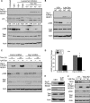
Inhibition of mTOR signaling by Dig2/RTP801/REDD1 in dexamethasone-treated WEHI7.2 cells. A, protein lysates of WEHI7.2 cells overexpressing Bcl-2 were collected following no treatment (untreated) or 4 h of treatment with vehicle (VEH, 0.1% ethanol), rapamycin (20 nm), or a dose response of dexamethasone in the presence of lysosomal inhibitors where indicated (pepstatin A, 10 μg/ml, and E64d, 5 μg/ml). Immunoblots were probed for Dig2/RTP801/REDD1, LC3, Thr-389-phosphorylated S6K (pS6K), and total S6K. The values represent normalized levels of LC3-II expression relative to Actin. B, Bcl-2-positive WEHI7.2 cells were transiently transfected with nontargeting control siRNA (NT) and Dig2/RTP801/REDD1 siRNA (KD), followed by treatment with vehicle (0.1% ethanol) or 1 μm dexamethasone for 4 h. mTOR activity was assessed by immunoblotting for Thr-389-phosphorylated S6K (pS6K) and total S6K. C, Bcl-2-positive WEHI7.2 cells stably transduced with lentiviral shRNA control and Dig2/RTP801/REDD1 shRNA were treated with vehicle (0.1% ethanol) or 1 μm dexamethasone for both 4 and 24 h. Immunoblotting was performed as in B. D, densitometry of immunoblot signal intensity from 4 h of treatments was used to quantify relative expression levels of pS6K. Signal intensity was normalized to the signal intensity of Actin. E, Bcl-2-positive WEHI7.2 cells were transiently transfected with 1000 nm nontargeting siRNA (NT) or TSC2 siRNA (KD) and after 16 h were treated with 1 μm dexamethasone or vehicle for 4 h prior to immunoblotting for Dig2/RTP801/REDD1, pS6K and total S6K. F, wild type WEHI7.2 cells were treated with 1 μm dexamethasone or vehicle for 4 h prior to immunoblotting for Dig2/RTP801/REDD1, pS6K, and total S6K. The error bars represent the means ± S.E. of at least three independent experiments. All of the immunoblots are representative of multiple independent experiments. *, p < 0.05 between control and Dig2/RTP801/REDD1 knockdown dexamethasone-treated samples.
FIGURE 5.
FIGURE 5.
Dig2/RTP801/REDD1 knock-out mouse thymocytes display partial inhibition of dexamethasone-induced autophagy. A–C, primary thymocytes were isolated from wild type and Dig2/RTP801/REDD1 knock-out mice following intraperitoneal injections of vehicle (VEH, phosphate-buffered saline) or dexamethasone (Dex) sodium phosphate for 4 h. The mice were treated with vehicle or 25 μg of dexamethasone sodium phosphate. A, immunoblots for Dig2/RTP801/REDD1 and LC3. B, the immunoblot signal for LC3-II was quantified using densitometry relative to actin and normalized to vehicle-treated mice for two independent experiments. C, Thr-389-phosphorylated S6K (pS6K) and total S6K. D, wild type and Dig2/RTP801/REDD1 knock-out mice were given intraperitoneal injections of vehicle or 200 μg of dexamethasone for 24 h. Primary thymocytes were isolated, and cell death was assayed using annexin V/propidium iodide staining and was quantified using flow cytometry. The error bars represent the means ± S.D. All of the immunoblots are representative of multiple independent experiments.

References

    1. Dougherty T. F., White A. (1943) Science 98, 367–369 - PubMed
    1. Hench P. S., Kendall E. C. (1949) Mayo Clin. Proc. 24, 181–197 - PubMed
    1. Pearson O. H., Eliel L. P. (1949) Cancer 2, 943–945 - PubMed
    1. Rhen T., Cidlowski J. A. (2005) N. Engl. J. Med. 353, 1711–1723 - PubMed
    1. Bowen D. A., Call T. G., Jenkins G. D., Zent C. S., Schwager S. M., Van Dyke D. L., Jelinek D. F., Kay N. E., Shanafelt T. D. (2007) Leuk. Lymphoma 48, 2412–2417 - PubMed

Publication types

MeSH terms

LinkOut - more resources