

Role of shear stress and stretch in vascular mechanobiology
- PMID: 21733876
- PMCID: PMC3163429
- DOI: 10.1098/rsif.2011.0177
Role of shear stress and stretch in vascular mechanobiology
Abstract
Blood vessels are under constant mechanical loading from blood pressure and flow which cause internal stresses (endothelial shear stress and circumferential wall stress, respectively). The mechanical forces not only cause morphological changes of endothelium and blood vessel wall, but also trigger biochemical and biological events. There is considerable evidence that physiologic stresses and strains (stretch) exert vasoprotective roles via nitric oxide and provide a homeostatic oxidative balance. A perturbation of tissue stresses and strains can disturb biochemical homeostasis and lead to vascular remodelling and possible dysfunction (e.g. altered vasorelaxation, tone, stiffness, etc.). These distinct biological endpoints are caused by some common biochemical pathways. The focus of this brief review is to point out some possible commonalities in the molecular pathways in response to endothelial shear stress and circumferential wall stretch.
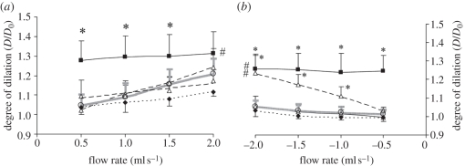
Figures

References
-
- Califano J. P., Reinhart-King C. A. 2010. Exogenous and endogenous force regulation of endothelial cell behavior. J. Biomech. 43, 79–86 10.1016/j.jbiomech.2009.09.012 (doi:10.1016/j.jbiomech.2009.09.012) - DOI - PubMed
-
- Agabiti-Rosei E., Rizzoni D. 2010. Regression of small resistance artery structural alterations in hypertension by appropriate antihypertensive treatment. Curr. Hypertens. Rep. 12, 80–85 10.1007/s11906-010-0093-7 (doi:10.1007/s11906-010-0093-7) - DOI - PubMed
-
- Sachidanandam K., Hutchinson J. R., Elgebaly M. M., Mezzetti E. M., Dorrance A. M., Motamed K., Ergul A. 2009. Glycemic control prevents microvascular remodeling and increased tone in type 2 diabetes: link to endothelin-1. Am. J. Physiol. Regulatory, Integr. Comp. Physiol. 296, R952–R959 10.1152/ajpregu.90537.2008 (doi:10.1152/ajpregu.90537.2008) - DOI - PMC - PubMed
-
- Creager M. A., Luscher T. F., Cosentino F., Beckman J. A. 2003. Diabetes and vascular disease: pathophysiology, clinical consequences, and medical therapy—part I. Circulation 108, 1527–1532 10.1161/01.CIR.0000091257.27563.32 (doi:10.1161/01.CIR.0000091257.27563.32) - DOI - PubMed
-
- Chien S. 2007. Mechanotransduction and endothelial cell homeostasis: the wisdom of the cell. Am. J. Physiol. Heart Circ. Physiol. 292, H1209–H1224 10.1152/ajpheart.01047.2006 (doi:10.1152/ajpheart.01047.2006) - DOI - PubMed
Publication types
MeSH terms
Grants and funding
LinkOut - more resources
Full Text Sources
Other Literature Sources
Medical