Review
. 2011 Oct 7;278(1720):2857-65.
doi: 10.1098/rspb.2011.0938. Epub 2011 Jul 6.

Evolution of ruminant headgear: a review

Affiliations
Review

Evolution of ruminant headgear: a review

Edward Byrd Davis et al. Proc Biol Sci. .

Abstract

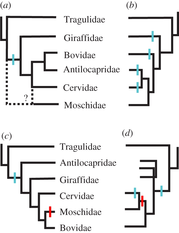
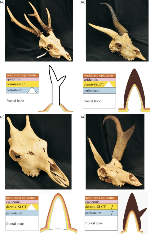
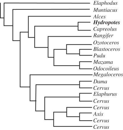
The horns, ossicones and antlers of ruminants are familiar and diverse examples of cranial appendages. We collectively term ruminant cranial appendages 'headgear'; this includes four extant forms: antlers (in cervids), horns (in bovids), pronghorns (in pronghorn antelope) and ossicones (in giraffids). Headgear evolution remains an open and intriguing question because phylogenies (molecular and morphological), adult headgear structure and headgear development (where data are available) all suggest different pictures of ruminant evolution. We discuss what is known about the evolution of headgear, including the evidence motivating previous hypotheses of single versus multiple origins, and the implications of recent phylogenetic revisions for these hypotheses. Inclusion of developmental data is critical for progress on the question of headgear evolution, and we synthesize the scattered literature on this front. The areas most in need of attention are early development in general; pronghorn and ossicone development in particular; and histological study of fossil forms of headgear. An integrative study of headgear development and evolution may have ramifications beyond the fields of systematics and evolution. Researchers in organismal biology, as well as those in biomedical fields investigating skin, bone and regenerative medicine, may all benefit from insights produced by this line of research.


Figures

Figure 1.
Figure 1.
History of phylogenetic hypotheses of ruminant family-level relationships, with superimposed hypotheses of character state transitions for headgear, as referred to in-text; gains are indicated in blue and losses in red. (a) Pilgrim [13], based on morphology, reflecting the putative homology of bovid and antilocaprid headgear and the uncertain position of Moschidae, hypothesized a single origin of headgear with no loss, now rejected. (b) Janis & Scott [14], based on morphology, but down-weighting cranial appendages, hypothesized four independent origins of headgear. (c) The topologies of both Marcot [9] and Spaulding et al. [10] suggest a single origin of headgear with one loss; the former study is based on a supermatrix of 16 different genetic markers; the latter is based on a combined evidence matrix of 12222 informative characters. (d) Hernández Fernández & Vrba [8], a supertree based on molecular and morphological data for extant ruminants, showing one example of an alternative hypothesis of headgear evolution, with two origins and one loss.
Figure 2.
Figure 2.
Ruminant headgear types discussed in this review. Upper photographs illustrate adult osteological forms. Lower left diagrams are simplified representations of the layers of tissue at the loci of headgear ossification during earliest ontogeny, with five tissue types illustrated with different coloured layers. From lowest (most internal) to highest (external), the layers are: white for the frontal bone, blue for the periosteum surrounding the frontal, yellow for the connective tissue (dermis + subcutaneous loose connective tissue (SLCT)), pink for the epidermis and brown for the keratinized epidermal sheath. A white triangle indicates the presumed site of initial ossification of headgear. Lower right diagrams illustrate tissues in adult states. (a) Cervid: Rusa unicolor University of California Museum of Vertebrate Zoology (MVZ) 181527; left diagram showing the extension of the frontal by a modified endochondral ossification; right diagram showing mature form composed primarily of exposed bone (integument and SLCT are shed by this stage). (b) Bovid: Antidorcas marsupialis MVZ 117913; left diagram showing the dermal origin of the horncore-forming os cornu or anlage, which either fuses with the frontal or stimulates outgrowth of bone; right diagram showing adult form composed mostly of bone and keratin, with two nested layers of keratin sheath indicating annual growth. (c) Giraffa camelopardalis MVZ 55148; left diagram showing initial dermal ossification; right diagram showing adult form composed of bone and skin. (d) Antilocapra americana MVZ 98089; left diagram indicating limited knowledge of early development, with suggestion of antler-like extension of frontal; right diagram showing mature form composed of bone, skin and keratin, with two centres of keratinization for each tine of the sheath.
Figure 3.
Figure 3.
Schematic of current genus-level cervid tree, with the antlerless Hydropotes highlighted in bold. Tree from Marcot [9].

References

    1. Jarman P. J. 1974. The social organisation of antelope in relation to their ecology. Behaviour 48, 215–26710.1163/156853974X00345 (doi:10.1163/156853974X00345) - DOI - DOI
    1. Reeves R. R., Tracey S. 1980. Monodon monoceros Mamm. Spec. 127, 1–710.2307/3503952 (doi:10.2307/3503952) - DOI - DOI
    1. Schneider D. 1964. Insect antennae. Annu. Rev. Entomol. 9, 103–12210.1146/annurev.en.09.010164.000535 (doi:10.1146/annurev.en.09.010164.000535) - DOI - DOI
    1. Krieger J., Breer H. 1999. Olfactory reception in invertebrates. Science 286, 720–72310.1126/science.286.5440.720 (doi:10.1126/science.286.5440.720) - DOI - DOI - PubMed
    1. Fay F. H. 1985. Odobenus rosmarus. Mamm. Spec. 238, 1–710.2307/3503810 (doi:10.2307/3503810) - DOI - DOI