. 2012 Jan;33(1):120-8.
doi: 10.1093/eurheartj/ehr196. Epub 2011 Jul 6.

Capture of circulatory endothelial progenitor cells and accelerated re-endothelialization of a bio-engineered stent in human ex vivo shunt and rabbit denudation model

Affiliations

Capture of circulatory endothelial progenitor cells and accelerated re-endothelialization of a bio-engineered stent in human ex vivo shunt and rabbit denudation model

Katarína Larsen et al. Eur Heart J. 2012 Jan.

Abstract

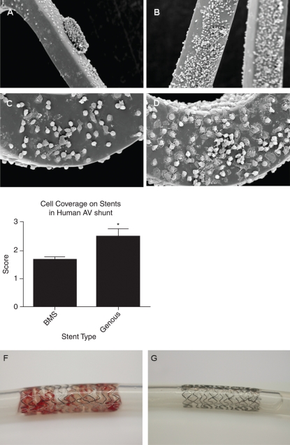
Aims: The Genous™ Bio-engineered R™ stent (GS) aims to promote vascular healing by capture of circulatory endothelial progenitor cells (EPCs) to the surface of the stent struts, resulting in accelerated re-endothelialization. Here, we assessed the function of the GS in comparison to bare-metal stent (BMS), when exposed to the human and animal circulation.

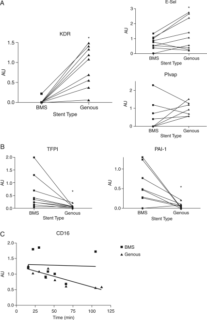
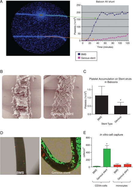
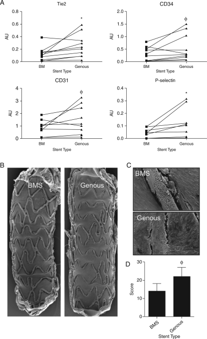
Methods and results: First, 15 patients undergoing coronary angiography received an extracorporeal femoral arteriovenous (AV) shunt containing BMS and GS. Macroscopical mural thrombi were observed in BMS, whereas GS remained visibly clean. Confocal and scanning electron microscopic (SEM) analysis of GS showed an increase in strut coverage. Quantitative polymerase chain reaction (qPCR) analysis of captured cells on the GS demonstrated increased expression of endothelial markers KDR/VEGFR2 and E-selectin, and a decrease in pro-thrombogenic markers tissue factor pathway inhibitor and plasminogen activator inhibitor-1 compared with BMS. Secondly, a similar primate AV shunt model was used to validate these findings and occlusion of BMS was observed, while GS remained patent, as demonstrated by live imaging of indium-labelled platelets. Thirdly, in an in vitro cell-capture assay, GS struts showed increased coverage by EPCs, whereas monocyte coverage remained similar to BMS. Finally, the assessment of re-endothelialization was studied in a rabbit denudation model. Twenty animals received BMS and GS in the aorta and iliac arteries for 7 days. Scanning electron microscopic analysis showed a trend towards increased strut coverage, confirmed by qPCR analysis revealing increased levels of endothelial markers (Tie2, CD34, PCD31, and P-selectin) in GS.

Conclusion: In this proof-of-concept study, we have demonstrated that the bio-engineered EPC-capture stent, Genous™ R™ stent, is effective in EPC capture, resulting in accelerated re-endothelialization and reduced thrombogenicity.


Figures

Figure 1
Figure 1
Scanning electron microscopic analysis of the endothelial progenitor cell capturing stent and bare-metal stent in the human arteriovenous shunt model revealed a marked increase in cell strut coverage compared with bare-metal stent. Scanning electron microscopic inspection of the study stents showed less strut coverage and the presence of thrombus-like structures on bare-metal stent (A) when compared with Genous stent (B). High-magnification scanning electron microscope revealed more adhesion of cells with a flattened polygonal morphology on the struts of the Genous stent (D) vs. bare-metal stent (C). Average stent-strut coverage was visually rated by blinded (core lab) technicians (CV-Path Institute, USA) on a 0–3 virtual scale (corresponding to 0–25, 25–50, 50–75, and 75–100% stent coverage). Bar graph indicates the level of strut coverage as assessed by scanning electron microscope in the two stent study groups. *P< 0.05, n = 9 (E). Macroscopic appearance of bare-metal stent (F) and Genous (G) stents in the human ex vivo shunt model shows mural thrombi in the bare-metal stent, whereas the Genous stent remained free of thrombogenic material.
Figure 2
Figure 2
Quantitative polymerase chain reaction evaluation of cellular markers in cell lysates of captured cells in the human arteriovenous shunt stent model. Paired comparison of the expression levels of the individual genes revealed a marked increase in endothelial markers, including KDR/VEGFR2 (P< 0.001) and E-selectin (P< 0.045) mRNA expression in the stents compared with bare-metal stent (A). Expression of another endothelial specific marker PLVAP (A) showed no significant increased expression in the stent (P = 0.21). Quantitative polymerase chain reaction analysis of markers of thrombosis, coagulation, and inflammation. Paired comparison of the expression levels of the individual genes revealed a marked decrease in tissue factor pathway inhibitor and plasminogen activator inhibitor-1 in the Genous compared with the bare-metal stent (B) (P = 0.04 and 0.02). Quantitative polymerase chain reaction showed a significant decrease in CD16 marker expression in the cells captured by the Genous stent over time, whereas the CD16 mRNA levels on bare-metal stent were maintained (C) (*P< 0.05).
Figure 3
Figure 3
Live imaging of arteriovenous shunt setup using a gamma camera to measure deposition of indium-labelled platelets on the study stents (A). Line graph shows a typical example of accumulating platelet signal over time. Low-magnification scanning electron microscope images of the bare-metal stent and the endothelial progenitor cell-capture stent in the baboon arteriovenous shunt model revealed a decrease in mural thrombus in the Genous vs. bare-metal stent (B). Bar graph showing the quantified number of platelets accumulated on the bare-metal stent and Genous stent after 2 h of flow exposure (C). Data were acquired from the live imaging of arteriovenous shunt setup, *P< 0.05, n = 3. In vitro assay to test the CD34+ cell-capture specificity of the Genous stent. Genous and BM stents were deployed in silicon tubing and were exposed to a cell mixture of PKH26 red fluorescent-labelled human monocytes (1 × 10⁶ cells/mL) and PKH2 green fluorescent-labelled human CD34+ cells (2 × 10⁵ cells/mL), under a constant rotation speed of 0.3 RPM for 2 h. Micrographs show confocal images of strut coverage of bare-metal stent and Genous stent (D). Bar graph shows the quantified number of CD34+ cells and monocytes per cm2 strut area. *P< 0.05, n = 3 (E).
Figure 4
Figure 4
Quantitative polymerase chain reaction analysis of the study stents of 11 New Zealand white rabbits was performed to evaluate capture of cells and subsequent expression of endothelial cell markers. Paired quantitative polymerase chain reaction analysis showed increased levels of endothelial markers by the cells captured on the Genous stent vs. bare-metal stent treated arteries, including Tie2 (P= 0.02), CD34 (P = 0.07), CD31 (P = 0.08), and P-selectin (P = 0.05). *P< 0.05, φP< 0.01, n = 11 (A). Scanning electron microscopic analysis of the stents implanted iliac vessels of 9 New Zealand white rabbits: Low (B)- and high (C)-magnification assessment revealed improved cell coverage between and above struts in the Genous stent vs. bare-metal stent. Bar graph shows the level of strut coverage as analysed by scanning electron microscope in the two stent groups. φP < 0.01, n = 9 (D).

References

    1. Urbich C, Dimmeler S. Endothelial progenitor cells: characterization and role in vascular biology. Circ Res. 2004;95:343–353. - PubMed
    1. Asahara T, Murohara T, Sullivan A. Isolation of putative progenitor endothelial cells for angiogenesis. Science. 1997;275:964–967. - PubMed
    1. Gulati R, Simari RD. Cell therapy for acute myocardial infarction. Med Clin North Am. 2007;91:769–785. xiii. - PubMed
    1. Hristov M, Erl W, Weber PC. Endothelial progenitor cells: mobilization, differentiation, and homing. Arterioscler Thromb Vasc Biol. 2003;23:1185–1189. - PubMed
    1. Kipshidze N, Dangas G, Tsapenko M. Role of the endothelium in modulating neointimal formation: vasculoprotective approaches to attenuate restenosis after percutaneous coronary interventions. J Am Coll Cardiol. 2004;44:733–739. - PubMed

Publication types

MeSH terms