Amino acid residues 253 and 591 of the PB2 protein of avian influenza virus A H9N2 contribute to mammalian pathogenesis
- PMID: 21734052
- PMCID: PMC3165745
- DOI: 10.1128/JVI.00702-11
Amino acid residues 253 and 591 of the PB2 protein of avian influenza virus A H9N2 contribute to mammalian pathogenesis
Abstract
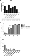
We investigated the tropism, host responses, and virulence of two variants of A/Quail/Hong Kong/G1/1997 (H9N2) (H9N2/G1) with D253N and Q591K in the PB2 protein in primary human macrophages and bronchial epithelium in vitro and in mice in vivo. Virus with PB2 D253N and Q591K had greater polymerase activity in minireplicon assays, induced more tumor necrosis factor alpha (TNF-α) in human macrophages, replicated better in differentiated normal human bronchial epithelial (NHBE) cells, and was more pathogenic for mice. Taken together, our studies help define the viral genetic determinants that contribute to pathogenicity of H9N2 viruses.
Figures


Similar articles
-
Assessment of the internal genes of influenza A (H7N9) virus contributing to high pathogenicity in mice.J Virol. 2015 Jan;89(1):2-13. doi: 10.1128/JVI.02390-14. Epub 2014 Oct 15. J Virol. 2015. PMID: 25320305 Free PMC article.
-
Virulence determinants in the PB2 gene of a mouse-adapted H9N2 virus.J Virol. 2015 Jan;89(1):877-82. doi: 10.1128/JVI.01775-14. Epub 2014 Oct 22. J Virol. 2015. PMID: 25339773 Free PMC article.
-
Double mutations in the H9N2 avian influenza virus PB2 gene act cooperatively to increase viral host adaptation and replication for human infections.J Gen Virol. 2021 Jun;102(6). doi: 10.1099/jgv.0.001612. J Gen Virol. 2021. PMID: 34061017
-
Adaptation of H9N2 Influenza Viruses to Mammalian Hosts: A Review of Molecular Markers.Viruses. 2020 May 14;12(5):541. doi: 10.3390/v12050541. Viruses. 2020. PMID: 32423002 Free PMC article. Review.
-
The role of the influenza virus RNA polymerase in host shut-off.Virulence. 2010 Sep-Oct;1(5):436-9. doi: 10.4161/viru.1.5.12967. Virulence. 2010. PMID: 21178485 Free PMC article. Review.
Cited by
-
Large-scale sequence analysis reveals novel human-adaptive markers in PB2 segment of seasonal influenza A viruses.Emerg Microbes Infect. 2018 Mar 29;7(1):47. doi: 10.1038/s41426-018-0050-0. Emerg Microbes Infect. 2018. PMID: 29593225 Free PMC article.
-
Anti-inflammatory effects of indirubin derivatives on influenza A virus-infected human pulmonary microvascular endothelial cells.Sci Rep. 2016 Jan 6;6:18941. doi: 10.1038/srep18941. Sci Rep. 2016. PMID: 26732368 Free PMC article.
-
Characterization in vitro and in vivo of a pandemic H1N1 influenza virus from a fatal case.PLoS One. 2013;8(1):e53515. doi: 10.1371/journal.pone.0053515. Epub 2013 Jan 10. PLoS One. 2013. PMID: 23326447 Free PMC article.
-
Transmission of influenza A viruses.Virology. 2015 May;479-480:234-46. doi: 10.1016/j.virol.2015.03.009. Epub 2015 Mar 24. Virology. 2015. PMID: 25812763 Free PMC article. Review.
-
An intermediate state allows influenza polymerase to switch smoothly between transcription and replication cycles.Nat Struct Mol Biol. 2023 Aug;30(8):1183-1192. doi: 10.1038/s41594-023-01043-2. Epub 2023 Jul 24. Nat Struct Mol Biol. 2023. PMID: 37488357
References
-
- Alexander D. J. 2007. An overview of the epidemiology of avian influenza. Vaccine 25:5637–5644 - PubMed
Publication types
MeSH terms
Substances
LinkOut - more resources
Full Text Sources

