

Implication of microRNAs in atrial natriuretic peptide and nitric oxide signaling in vascular smooth muscle cells
- PMID: 21734186
- PMCID: PMC3191566
- DOI: 10.1152/ajpcell.00088.2011
Implication of microRNAs in atrial natriuretic peptide and nitric oxide signaling in vascular smooth muscle cells
Abstract
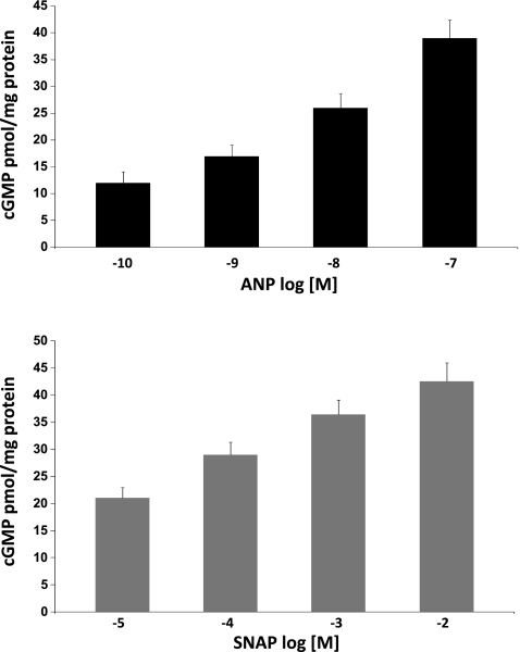
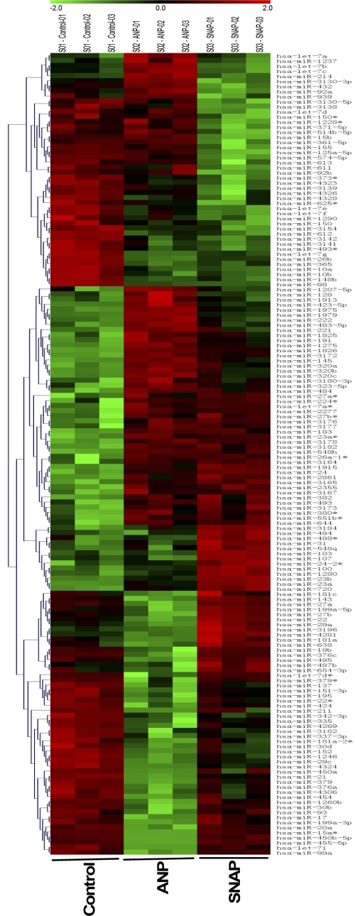
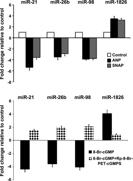
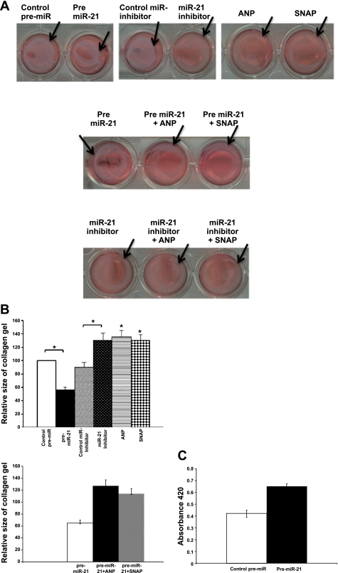
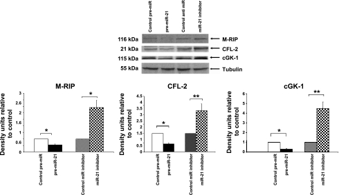
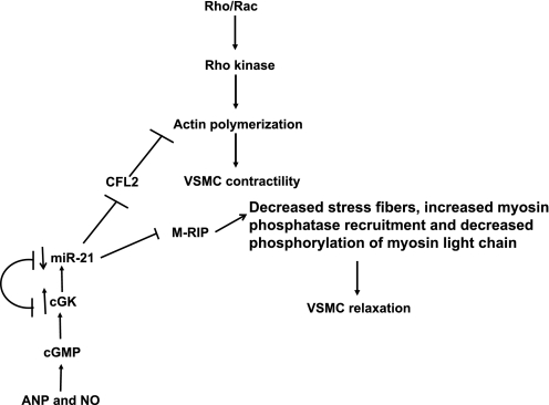
MicroRNAs (miRs) are endogenous small RNA molecules that suppress gene expression by binding to complementary sequences in the 3' untranslated regions of their target genes. miRs have been implicated in many diseases, including heart failure, ischemic heart disease, hypertension, cardiac hypertrophy, and cancers. Nitric oxide (NO) and atrial natriuretic peptide (ANP) are potent vasorelaxants whose actions are mediated through receptor guanylyl cyclases and cGMP-dependent protein kinase. The present study examines miRs in signaling by ANP and NO in vascular smooth muscle cells. miR microarray analysis was performed on human vascular smooth muscle cells (HVSMC) treated with ANP (10 nM, 4 h) and S-nitroso-N-acetylpenicillamine (SNAP) (100 μM, 4 h), a NO donor. Twenty-two shared miRs were upregulated, and 21 shared miRs were downregulated, by both ANP and SNAP (P < 0.05). Expression levels of four miRs (miRs-21, -26b, -98, and -1826), which had the greatest change in expression, as determined by microarray analysis, were confirmed by quantitative RT-PCR. Rp-8-Br-PET-cGMPS, a cGMP-dependent protein kinase-specific inhibitor, blocked the regulation of these miRs by ANP and SNAP. 8-bromo-cGMP mimicked the effect of ANP and SNAP on their expression. miR-21 was shown to inhibit HVSMC contraction in collagen gel lattice contraction assays. We also identified by computational algorithms and confirmed by Western blot analysis new intracellular targets of miR-21, i.e., cofilin-2 and myosin phosphatase and Rho interacting protein. Transfection with pre-miR-21 contracted cells and ANP and SNAP blocked miR-21-induced HVSMC contraction. Transfection with anti-miR-21 inhibitor reduced contractility of HVSMC (P < 0.05). The present results implicate miRs in NO and ANP signaling in general and miR-21 in particular in cGMP signaling and vascular smooth muscle cell relaxation.
Figures
Similar articles
-
Natriuretic peptides and nitric oxide stimulate cGMP synthesis in different cellular compartments.J Gen Physiol. 2006 Jul;128(1):3-14. doi: 10.1085/jgp.200509403. Epub 2006 Jun 12. J Gen Physiol. 2006. PMID: 16769793 Free PMC article.
-
Nitric oxide, atrial natriuretic peptide, and cyclic GMP inhibit the growth-promoting effects of norepinephrine in cardiac myocytes and fibroblasts.J Clin Invest. 1998 Feb 15;101(4):812-8. doi: 10.1172/JCI119883. J Clin Invest. 1998. PMID: 9466976 Free PMC article.
-
Regulation of cGMP by soluble and particulate guanylyl cyclases in cultured human airway smooth muscle.Am J Physiol. 1997 Oct;273(4):L807-13. doi: 10.1152/ajplung.1997.273.4.L807. Am J Physiol. 1997. PMID: 9357856
-
Regulation of vascular smooth muscle cell phenotype by cyclic GMP and cyclic GMP-dependent protein kinase.Front Biosci. 2006 Jan 1;11:356-67. doi: 10.2741/1803. Front Biosci. 2006. PMID: 16146737 Review.
-
Nutrition, microRNAs, and Human Health.Adv Nutr. 2017 Jan 17;8(1):105-112. doi: 10.3945/an.116.013839. Print 2017 Jan. Adv Nutr. 2017. PMID: 28096131 Free PMC article. Review.
Cited by
-
Investigation of Antihypertensive Properties of Chios Mastic via Monitoring microRNA-21 Expression Levels in the Plasma of Well-Controlled Hypertensive Patients.Noncoding RNA. 2024 May 31;10(3):33. doi: 10.3390/ncrna10030033. Noncoding RNA. 2024. PMID: 38921830 Free PMC article.
-
DiGeorge syndrome critical region 8 (DGCR8) protein-mediated microRNA biogenesis is essential for vascular smooth muscle cell development in mice.J Biol Chem. 2012 Jun 1;287(23):19018-28. doi: 10.1074/jbc.M112.351791. Epub 2012 Apr 17. J Biol Chem. 2012. PMID: 22511778 Free PMC article.
-
Hypertension: Potential Player in Cardiovascular Disease Incidence in Preeclampsia.Cardiovasc Toxicol. 2022 May;22(5):391-403. doi: 10.1007/s12012-022-09734-w. Epub 2022 Mar 28. Cardiovasc Toxicol. 2022. PMID: 35347585 Review.
-
MicroRNA and Heart Failure.Int J Mol Sci. 2016 Apr 6;17(4):502. doi: 10.3390/ijms17040502. Int J Mol Sci. 2016. PMID: 27058529 Free PMC article. Review.
-
MicroRNAs in hypertension: mechanisms and therapeutic targets.Curr Hypertens Rep. 2012 Feb;14(1):79-87. doi: 10.1007/s11906-011-0235-6. Curr Hypertens Rep. 2012. PMID: 22052337 Review.
References
-
- Albinsson S, Nordstrom I, Hellstrand P. Stretch of the vascular wall induces smooth muscle differentiation by promoting actin polymerization. J Biol Chem 279: 34849–34855, 2004 - PubMed
Publication types
MeSH terms
Substances
Grants and funding
LinkOut - more resources
Full Text Sources