. 2011 Sep 1;118(9):2551-5.
doi: 10.1182/blood-2010-12-324707. Epub 2011 Jul 6.

Inhibition of TET2-mediated conversion of 5-methylcytosine to 5-hydroxymethylcytosine disturbs erythroid and granulomonocytic differentiation of human hematopoietic progenitors

Affiliations

Inhibition of TET2-mediated conversion of 5-methylcytosine to 5-hydroxymethylcytosine disturbs erythroid and granulomonocytic differentiation of human hematopoietic progenitors

Elodie Pronier et al. Blood. .

Abstract

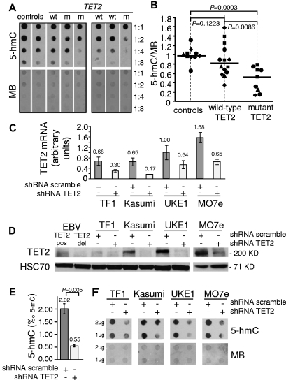
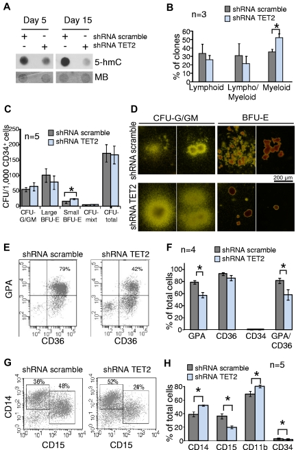
TET2 converts 5-methylcytosine to 5-hydroxymethylcytosine (5-hmC) in DNA and is frequently mutated in myeloid malignancies, including myeloproliferative neoplasms. Here we show that the level of 5-hmC is decreased in granulocyte DNA from myeloproliferative neoplasm patients with TET2 mutations compared with granulocyte DNA from healthy patients. Inhibition of TET2 by RNA interference decreases 5-hmC levels in both human leukemia cell lines and cord blood CD34(+) cells. These results confirm the enzymatic function of TET2 in human hematopoietic cells. Knockdown of TET2 in cord blood CD34(+) cells skews progenitor differentiation toward the granulomonocytic lineage at the expense of lymphoid and erythroid lineages. In addition, by monitoring in vitro granulomonocytic development we found a decreased granulocytic differentiation and an increase in monocytic cells. Our results indicate that TET2 disruption affects 5-hmC levels in human myeloid cells and participates in the pathogenesis of myeloid malignancies through the disturbance of myeloid differentiation.


Figures

Figure 1
Figure 1
Mutation or knockdown of TET2 lead to decreased levels of 5-hmC in human hematopoietic cells. (A) 5-hmC content of granulocytes from healthy patients (controls) and wild-type (wt) or mutant (m) TET2 MPN patients was determined by analyzing serial 2-fold dilutions of granulocyte DNA on dot blots with an anti–5-hmC antibody. The membranes were stained by MB to allow quantification of spot intensities by ImageJ software. (B) The ratios of 5-hmC to MB spot intensities from 4 experiments (10 control samples, 14 wild-type TET2 MPN samples, and 9 mutant TET2 MPN samples) were calculated, and individual values were normalized to the average value of control DNA for each experiment. Normalized values are plotted as squares, triangles, diamonds, and circles, each symbol representing one experiment. Horizontal bars indicate median values. P values were obtained with the use of the 2-tailed Mann-Whitney test. (C) TET2 mRNA quantification in TF1, Kasumi-1, UKE1, and MO7e cell lines transduced by lentiviruses expressing GFP and shRNA designed against either TET2 (shRNA TET2) or a control scramble sequence (shRNA scramble). Error bars indicate SEM. (D) Western blot analysis of transduced cell lines with anti-TET2 and anti-HSC70 antibodies. EBV lymphoblastoid cell lines from a patient with no TET2 mutation (EBV TET2 pos) and a patient with a biallelic deletion of TET2 (EBV TET2 del) were used as positive and negative controls. (E) 5-hmC content was determined by HPLC-MSMS in transduced MO7E cells. Histograms show 5-hmC/5-mC ratios (mean of 3 experiments). Error bars indicate SEM. P value was obtained with an unpaired Student t test. (F) A total of 1-2 μg of DNA from transduced TF1, Kasumi-1, UKE1, and MO7e cells was spotted for 5-hmC dot blot assay. Membranes were stained with MB to control spotting.
Figure 2
Figure 2
Knockdown of TET2 disturbs myeloid differentiation of cord blood CD34+ cells in vitro. Cord blood CD34+ cells were transduced by lentiviruses expressing GFP and either shRNA scramble or shRNA TET2. GFP+ cells were sorted 2 days after the end of the transduction procedure. (A) Sorted GFP+ cells were grown in MEM-α medium supplemented with SCF, FLT3-L, IL-3, and G-CSF. After 5-15 days of culture cells were harvested, DNA extracted, and spotted for 5-hmC dot blot assay. Membranes were stained with MB to assess equal spotting. Results are representative of 3 experiments, each performed at 3 time points of culture. (B) Sorted GFP+ CD34+CD38 cells were seeded at one cell per well on a confluent layer of MS-5 cells in a specific medium supporting B/NK/GM differentiation. After 4-6 weeks of culture wells with significant cell growth were harvested and 450 clones from 3 independent experiments were tested for B, natural killer, and myeloid differentiations with the use of anti-CD19, anti-CD56, and anti-CD15 antibodies. Histograms show the percentages of lymphoid (CD15 and CD56+ or CD19+), lympho/myeloid (CD15+CD19+ or CD15+CD56+ or CD15+CD19+CD56+), and myeloid (CD15+CD19CD56) clones. *P < .05, unpaired Student t test. Error bars indicate SEM. (C) Transduced CD34+ cells were seeded in methylcellulose in the presence of EPO, IL-3, SCF, and G-CSF. At day 14, colonies derived from shRNA scramble and shRNA TET2 BFU-E and CFU-G/GM were enumerated. Histograms show the number of colonies derived from transduced CD34+ cells (n = 5 independent experiments). *P < .05, unpaired Student t test. Error bars indicate SEM. (D) Photographs showing BFU-E– and CFU-G/GM–derived colonies. Colonies derived from shRNA TET2 BFU-E were smaller than those derived from shRNA scramble BFU-E. In contrast, CFU-G/GM–derived colonies appeared larger. (E) Transduced CD34+ cells were grown in serum-free medium supplemented with SCF, IL-3, and EPO. At day 14, CD34, CD36, and glycophorin-A (GPA) expression were analyzed by flow cytometry. Scattergrams from one representative of 4 experiments are shown. (F) Histograms represent the mean percentages of cells positive for GPA, CD36, CD34, and double-positive for GPA and CD36 antigens in the whole cell suspension after the 14-day culture. *P < .05, unpaired Student t test. Error bars indicate SEM (G) Transduced CD34+ cells were grown in a medium containing SCF, FLT3-L, IL-3, and G-CSF to monitor granulomonocytic differentiation in vitro. CD14 and CD15 immunophenotypic analysis at day 10 in 1 representative of 5 independent experiments is shown. (H) CD14, CD15, CD11b, and CD34 immunophenotypic analysis of cultured cells at day 10. Histograms represent the mean percentages of cells positive for each antigen in the whole cell suspension (5 independent experiments). *P < .05, unpaired Student t test. Error bars indicate SEM.

Comment in

References

    1. Tahiliani M, Koh KP, Shen Y, et al. Conversion of 5-methylcytosine to 5-hydroxymethylcytosine in mammalian DNA by MLL partner TET1. Science. 2009;324(5929):930–935. - PMC - PubMed
    1. Ito S, D'Alessio AC, Taranova OV, Hong K, Sowers LC, Zhang Y. Role of Tet proteins in 5mC to 5hmC conversion, ES-cell self-renewal and inner cell mass specification. Nature. 2010;466(7310):1129–1133. - PMC - PubMed
    1. Ficz G, Branco MR, Seisenberger S, et al. Dynamic regulation of 5-hydroxymethylcytosine in mouse ES cells and during differentiation. Nature. 2011;473(7347):398–402. - PubMed
    1. Tefferi A, Lim KH, Abdel-Wahab O, et al. Detection of mutant TET2 in myeloid malignancies other than myeloproliferative neoplasms: CMML, MDS, MDS/MPN and AML. Leukemia. 2009;23(7):1343–1345. - PMC - PubMed
    1. Langemeijer SM, Kuiper RP, Berends M, et al. Acquired mutations in TET2 are common in myelodysplastic syndromes. Nat Genet. 2009;41(7):838–842. - PubMed

Publication types

MeSH terms