Review
. 2011 Jul;24(3):459-68.
doi: 10.1128/CMR.00057-10.

Onchocerciasis: the role of Wolbachia bacterial endosymbionts in parasite biology, disease pathogenesis, and treatment

Affiliations
Review

Onchocerciasis: the role of Wolbachia bacterial endosymbionts in parasite biology, disease pathogenesis, and treatment

Francesca Tamarozzi et al. Clin Microbiol Rev. 2011 Jul.

Abstract

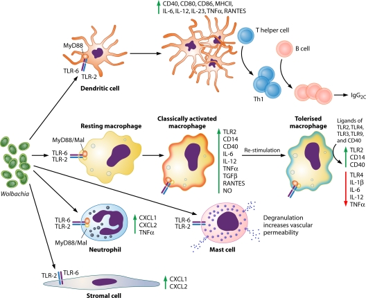
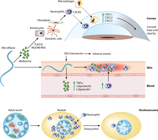
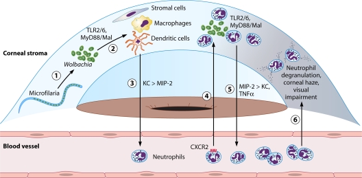
The discovery of Wolbachia intracellular bacteria within filarial nematodes, including Onchocerca volvulus, the causative agent of onchocerciasis or "river blindness," has delivered a paradigm shift in our understanding of the parasite's biology, to where we now know that the bacterial endosymbionts are essential for normal development of larvae and embryos and may support the long-term survival of adult worms. The apparent mutualistic dependency has also offered a novel approach to the treatment of onchocerciasis through the use of antibiotics to eliminate Wolbachia, delivering for the first time a treatment which has significant macrofilaricidal efficacy. Studies with other filarial nematode species have also highlighted a role for Wolbachia in transmission and infection of the mammalian host through a fascinating manipulation of mast cell-mediated vasodilation to enhance infectivity of vector-borne larvae. Wolbachia has also been identified as the principal driver of innate and adaptive Th1 inflammatory immunity, which can either contribute to disease pathogenesis or, with the Wolbachia-mediated recruitment of mast cells, enhance infectivity. The Wolbachia activation of innate inflammation also drives inflammatory adverse events in response to chemotherapy with either diethylcarbamazine (DEC) or ivermectin. In this review we summarize the experimental and field trial data which have uncovered the importance of Wolbachia symbiosis in onchocerciasis.


Figures

Fig. 1.
Fig. 1.
Wolbachia-induced responses of specific cell types. Wolbachia-exposed dendritic cells are activated via the TLR2/6-MyD88-Mal pathway, as shown by enhanced expression of surface costimulatory molecules and produce proinflammatory cytokines, inducing a preferential type 1 (Th1) immune response (11, 77). Macrophages stimulated with Wolbachia or Wolbachia-containing but not Wolbachia-depleted filarial extracts enhance their surface expression of costimulatory molecules and produce proinflammatory cytokines and oxidative products. Macrophages can be homo- and heterotolerized by a subsequent stimulation, contributing to the immune downregulation characterizing the majority of filarial infections (29, 67, 76, 77). Neutrophils and corneal stromal cells are also able to interact with Wolbachia via the TLR2-MyD88 pathway, producing CXC chemokines and contributing to the inflammatory response to the parasite (–24). Mast cells are stimulated by Wolbachia via TLR2 to degranulate and increase vascular permeability to facilitate establishment of infection (63). Abbreviations: TLR, Toll-like receptor; CD, cluster of differentiation; MHCII, major histocompatibility complex class II; IL, interleukin; TNF-α, tumor necrosis factor alpha; RANTES, regulated upon activation, normal T-cell expressed, and secreted; TGF-β, transforming growth factor β; NO, nitric oxide; MyD88, myeloid differentiation primary response gene (88); Mal, MyD88 adaptor-like.
Fig. 2.
Fig. 2.
Role of Wolbachia in river blindness. Wolbachia release after microfilaria death in the cornea causes corneal edema and opacity by inducing neutrophil and macrophage infiltration and activation in the corneal stroma that are dependent on TLR2-MyD88 activation and production of CXC chemokines. Keratocytes and bone marrow-derived cells in the corneal stroma can initiate this response, which is then perpetuated by inflammatory cells (–24, 29, 56, 77). When large loads of Wolbachia bacteria are released from microfilariae after microfilaricidal treatment, this induces cutaneous and systemic side effects such as fever, tachycardia, hypotension, lymphadenopathy, and pruritus. In the skin, neutrophils are the first cells to be recruited and activated, inducing dermal inflammation. At a systemic level, adverse events correlate with microfilarial loads and are associated with Wolbachia DNA and whole bacterial levels in blood, proinflammatory cytokines, neutrophilia, and antibacterial peptides (calprotectin and calgranulin) (25, 43, 52, 80). The presence of Wolbachia is associated with neutrophil infiltration in the cornea, skin, and onchocercomas (8, 22, 23). Abbreviations: TLR, Toll-like receptor; MyD88, myeloid differentiation primary response gene (88); TNF-α, tumor necrosis factor alpha; DEC, diethylcarbamazine.
Fig. 3.
Fig. 3.
Predicted sequence of events in O. volvulus/Wolbachia-induced keratitis. Wolbachia released from dying microfilariae in the corneal stroma (1) activates resident cells, including fibroblasts, dendritic cells, and macrophages (2). These cells produce chemokines MIP-2 and predominantly KC (3). KC induces a CXCR2-dependent neutrophil migration to the corneal stroma, where neutrophils are also activated via TLR2/6 by Wolbachia (4). Neutrophils produce additional chemokines, predominantly MIP-2, inducing further neutrophil migration (5). Neutrophils degrade the corneal matrix, causing corneal haze and visual impairment (6). Abbreviations: TLR, Toll-like receptor; MyD88, myeloid differentiation primary response gene (88); Mal, MyD88 adaptor-like; KC, keratinocyte-derived chemokine; MIP, macrophage-inflammatory protein 2; TNF-α, tumor necrosis factor alpha; DC, dendritic cell.

References

    1. Ali M. M., et al. 2003. Immune responses directed against microfilariae correlate with severity of clinical onchodermatitis and treatment history. J. Infect. Dis. 187:714–717 - PubMed
    1. Ali M. M., et al. 2007. Fc gamma RIIa (CD32) polymorphism and onchocercal skin disease: implications for the development of severe reactive onchodermatitis (ROD). Am. J. Trop. Med. Hyg. 77:1074–1078 - PubMed
    1. Baker R. H., Abdelnur O. M. 1986. Onchocerciasis in Sudan: the distribution of the disease and its vectors. Trop. Med. Parasitol. 37:341–355 - PubMed
    1. Basanez M. G., et al. 2006. River blindness: a success story under threat? PLoS Med. 3:e371. - PMC - PubMed
    1. Bennuru S., et al. 2009. Brugia malayi excreted/secreted proteins at the host/parasite interface: stage- and gender-specific proteomic profiling. PLoS Negl. Trop. Dis. 3:e410. - PMC - PubMed

MeSH terms