Review
. 2011 Jul;24(3):592-630.
doi: 10.1128/CMR.00063-10.

Pathogenesis of chagas' disease: parasite persistence and autoimmunity

Affiliations
Review

Pathogenesis of chagas' disease: parasite persistence and autoimmunity

Antonio R L Teixeira et al. Clin Microbiol Rev. 2011 Jul.

Abstract

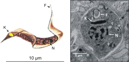
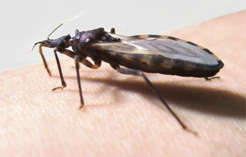
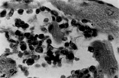
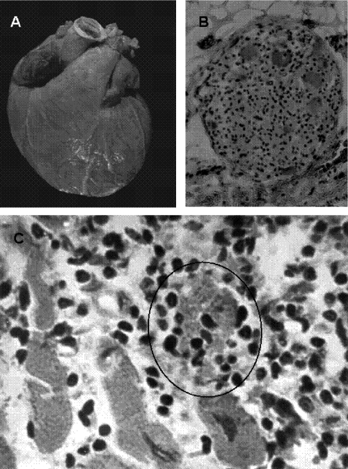
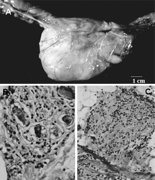
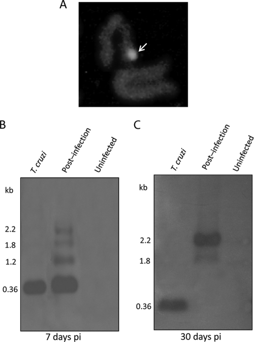
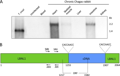
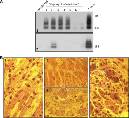
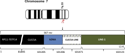
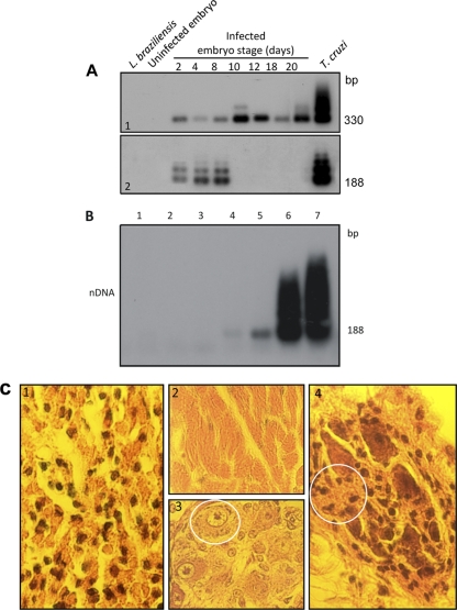
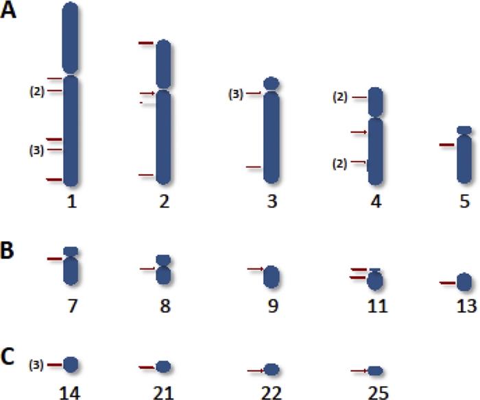
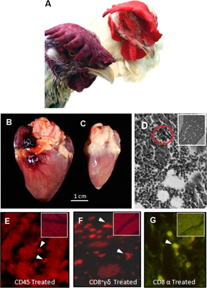
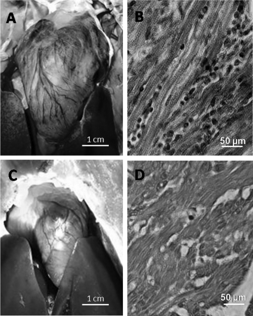
Acute Trypanosoma cruzi infections can be asymptomatic, but chronically infected individuals can die of Chagas' disease. The transfer of the parasite mitochondrial kinetoplast DNA (kDNA) minicircle to the genome of chagasic patients can explain the pathogenesis of the disease; in cases of Chagas' disease with evident cardiomyopathy, the kDNA minicircles integrate mainly into retrotransposons at several chromosomes, but the minicircles are also detected in coding regions of genes that regulate cell growth, differentiation, and immune responses. An accurate evaluation of the role played by the genotype alterations in the autoimmune rejection of self-tissues in Chagas' disease is achieved with the cross-kingdom chicken model system, which is refractory to T. cruzi infections. The inoculation of T. cruzi into embryonated eggs prior to incubation generates parasite-free chicks, which retain the kDNA minicircle sequence mainly in the macrochromosome coding genes. Crossbreeding transfers the kDNA mutations to the chicken progeny. The kDNA-mutated chickens develop severe cardiomyopathy in adult life and die of heart failure. The phenotyping of the lesions revealed that cytotoxic CD45, CD8(+) γδ, and CD8α(+) T lymphocytes carry out the rejection of the chicken heart. These results suggest that the inflammatory cardiomyopathy of Chagas' disease is a genetically driven autoimmune disease.


Figures

Fig. 1.
Fig. 1.
Trypanosoma cruzi parasitic forms present in human tissues. (Left) Schematic representation of a trypomastigote form of Trypanosoma cruzi. (Reprinted from reference with permission of the publisher.) (Right) Ultrastructure of a T. cruzi amastigote free in the cytoplasm of a muscle cell. N, nucleus; K, kinetoplast; F, flagellum.
Fig. 2.
Fig. 2.
Triatoma infestans (Hemiptera: reduviid), the main transmitter of Trypanosoma cruzi to humans. The adult kissing bug inserts its stylet into the skin for blood feeding from the forearm.
Fig. 3.
Fig. 3.
Trypanosoma cruzi amastigote nest in the heart of a naturally infected baboon. (A) Parasite nest (arrow) seen in a healthy myocardium section. Shown is hematoxylin and eosin (HE) staining. Magnification, ×200. (B) T. cruzi amastigotes fluorescing with a fluorescein-conjugated streptavidin 188-nt probe, which was PCR amplified with nDNA primers. The amastigote parasites hibernate in the nest in the absence of an inflammatory reaction in their surroundings. (Reprinted from reference with permission of the publisher.)
Fig. 4.
Fig. 4.
Pathology of acute human Chagas' disease. (A) Heart section with a nest of T. cruzi amastigote forms (arrows) and mononuclear cell infiltrates associated with lysis of parasite-free target myofibers. (B) Skeletal muscle showing inflammatory mononuclear cell infiltrates and target cell destruction. (C) Nodular inflammatory lesion in the gray matter of the brain. (Reprinted from reference with permission of the publisher.)
Fig. 5.
Fig. 5.
Heart lesion in the chronic human intermediate form. The minimal rejection unit consists of the lysis of the target myofiber by immune system mononuclear cells. (Reprinted from reference with permission of the publisher.)
Fig. 6.
Fig. 6.
Pathology of chronic human Chagas' heart disease. (A) Cardiomegaly in an adult with increased ventricles, bulging pulmonary artery conus, lymphatic vessel engorgement, and whitish soldier's patch. (B) Parasympathetic ganglion showing ganglionitis and neuronolysis. (C) Histopathological lesion consisting of strike mononuclear cell infiltrates and diffuse target heart cell lysis. A typical minimal rejection unit is encircled. (Reprinted from reference with permission of the publisher.)
Fig. 7.
Fig. 7.
Pathology of the megacolon in a chronic chagasic baboon. (A) Huge dilation and thickening of the walls of a segment of sigmoid colon and rectum. (B) Parasympathetic ganglionitis and neuronitis and loss of neurons. (C) Section of a sympathetic nerve on the serosa surface of the colon showing peri- and intraneuritis. (Reprinted from reference with permission of the publisher.)
Fig. 8.
Fig. 8.
Insertion of the Trypanosoma cruzi kDNA minicircle into the human macrophage genome. (A) Identification of the kDNA in a metaphase plate chromosome by the FISH method. Fluorescence (arrow) is seen in a chromosome probed with a biotin-labeled minicircle. (Reprinted from reference with permission of the publisher.) (B) Southern hybridization of NsiI digests of 7-day-postinfection (pi) macrophages with a kDNA probe on blots of 0.8% agarose gels. The T. cruzi kDNA minicircle forms a single 0.36-kb band, whereas the early-infection macrophage DNAs show upper 1.2-, 1.8-, and 2.2-kb bands. (Reprinted from reference with permission of the publisher.) (C) Hybridization of NsiI digests of 30-day (postinfection) macrophages with a kDNA probe. The absence of the 0.36-kb band in the Southern blot and the presence of the 1.8- and 2.2-kb PCR products in the 30-day-postinfection macrophage DNA indicate that the T. cruzi infection is eradicated and that the minicircle is inserted into the host cell genome. (Reprinted from reference with permission of the publisher.)
Fig. 9.
Fig. 9.
Integration of the kDNA minicircle sequence into the genome of a rabbit with Chagas' disease. (A) Hybridization of rabbit DNA with a specific kDNA probe. A total of 20 μg of EcoRI-digested DNA separated on a 0.7% agarose gel was used for Southern hybridization with 1 μg of a T. cruzi cloned kDNA constant region (kCR) probe. (B) Schematic representation of kDNA integration into rabbit DNA. Arrows show primers used for 5′ RACE to detect the kDNA insertion into the rabbit genomic clone LBNL1. The integration of CCA/ACC-rich kDNA (bp 1255 to 1907) occurred within rabbit DNA, showing attachment sites of direct short CACCAACC repeats. An ORF spans the chimeric sequence at bp 1217 to 1582. (Reprinted from reference with permission of the publisher [see comments regarding this retracted article at first citation].)
Fig. 10.
Fig. 10.
Genetic markers of T. cruzi infection in offspring of rabbits with Chagas' disease with evident pathology. (A) Specific hybridization of PCR amplification products from template DNA obtained from offspring of doe C with Chagas' disease using specific sets of T. cruzi nDNA and kDNA primers. DNA products were resolved on 1% agarose gels. (1) Analysis of kDNA amplification showing bands of 330 bp and its catemers from parasite DNA and from genomic DNA of six progeny with hybridization with the kCR probe. (2) Analysis of nDNA amplification showing bands of 188 bp and its catemers formed with parasite DNA and from genomic DNA of offspring 2 after hybridization with the specific internal probe. (B) Destructive myocarditis and ganglionitis present in 2-week-old offspring. (1) Histopathological section showing mononuclear cell infiltration and lysis of target heart cells. Note the round lymphocytes adhering to the surface of the target cells. (2) Normal histological features of myocardium. (3) Intracardiac ganglion cells from a control offspring of a noninfected rabbit. The circle depicts a neuron. (4) Intracardiac parasympathetic ganglion showing mononuclear cell infiltration and neuron dropout (circle). (Reprinted from reference with permission of the publisher [see comments regarding this retracted article at first citation].)
Fig. 11.
Fig. 11.
Topology of the Trypanosoma cruzi kDNA minicircle integrated into the genome of a Chagas' case and knockout of the CLEC5A gene. The scheme shows kDNA integration into the chromosome 7 CLEC5A gene open reading frame in continuity with LINE-1 at RP11-707F14. The tpTAIL-PCR amplicon that was cloned and sequenced shows that the s67 reverse kDNA primer anneals at the 5′ end and that the L1-5 primer anneals at the 3′ end. The kDNA (light blue, variable region) extends from nucleotides 1 to 286, and the host DNA (green) extends from nucleotides 270 to 1230. E values for kDNA and for host DNA are highly statistically significant. (Scheme reproduced from reference with permission of the publisher.)
Fig. 12.
Fig. 12.
Trypanosoma cruzi infection is established in a Gallus gallus embryo. (A) Positive-control T. cruzi trypomastigote fluorescein labeled with antibody from a Chagas' disease patient. The inset shows a fluorescein-labeled amastigote stained with Chagas' disease patient antibody diluted 1:128 in PBS (pH 7.4). (B) Section (HE stained) (magnification, ×100) from an uninfected chicken embryo which remained unstained with the anti-T. cruzi antibody diluted 1:128. (C) Background staining of the same control section treated with fluorescein-labeled anti-T. cruzi antibody. (D) Infected chicken embryo endoderm and mesoderm parasitized cells stained green by the fluorescein-labeled anti-T. cruzi antibody diluted 1:128. (E) X-gal (5-bromo-4-chloro-3-indolyl-β-d-galactopyranoside) blue-stained β-galactosidase-expressing T. cruzi Tulahuen-infected chicken embryo endoderm and mesoderm cells. Control serum from an uninfected donor lacking anti-T. cruzi antibody did not stain parasitized embryo cells. (Reprinted from PLoS Neglected Tropical Diseases [402].)
Fig. 13.
Fig. 13.
Evidence of kDNA integration into germ line cells and tissues from birds hatched from T. cruzi-infected eggs, with accompanying pathology. (A) Establishment of T. cruzi infection early in embryonic development followed by elimination early in Gallus gallus embryonic development. (Top) Bands (330 bp) formed by PCR-amplified minicircle kDNA templates harvested at several stages of chicken embryonic development, after hybridization with a specific probe. (Bottom) Bands formed by PCR amplified from the same embryos after separation on a 1% agarose gel and hybridization with a specific nDNA probe. The 188-bp nDNA band is diagnostic of the parasite persistence in the host tissue. (B) Sensitivity of PCR with nDNA primers Tcz1 and Tcz2. Lanes 1 and 2, control DNA from kDNA-negative and from kDNA-mutated chickens; lanes 3 to 7, mix of 200 ng of control chicken DNA with increasing amounts of T. cruzi DNA, 1 fg, 10 fg, 1 pg, 100 pg, and 1 ng, respectively. Hybridization with the radiolabeled 188-bp probe improved the sensitivity of the technique (10 fg), which is 24-fold below that for single T. cruzi total DNA. (C) Destructive myocarditis and ganglionitis in a 2-week-old F1 chick. (1) Histopathological section showing mononuclear cell infiltration and lysis of target heart cells. (2) Normal histological features of the myocardium. (3) Intracardiac ganglion cells from a control offspring of a noninfected chick. (4) Section of an intracardiac parasympathetic ganglion showing lymphocytic infiltration and dropout (circle) of neuronal cells. (Modified from reference with permission of the publisher [see comments regarding this retracted article at first citation] and from PLoS Neglected Tropical Diseases [402].)
Fig. 14.
Fig. 14.
Heredity of integrations of Trypanosoma cruzi kDNA minicircles into several loci of the chicken genome. Rows A, B, and C show integrations into the macrochromosomes, into the intermediate chromosomes, and into the microchromosomes, respectively. The numbers in parentheses indicate the total numbers of times that an insertion (red bar) was present at a chromosomal locus from animal source. (Reprinted from PLoS Neglected Tropical Diseases [402].)
Fig. 15.
Fig. 15.
Clinical and pathological findings for Gallus gallus with Trypanosoma cruzi kDNA mutations. (A) Nine-month-old F1 hen with heart insufficiency, showing cyanosis of the comb, and a control hen of the same age, showing a bright red comb. (B) Cardiomegaly (30 g) in a 9-month-old hen that died of heart failure. (C) Control heart (8 g) from a 9-month-old hen. (D) Myocarditis showing immune system mononuclear cell infiltrates and lysis of target heart cells. The red circle depicts a minimal rejection unit, whereby effector lymphocytes destroy a target heart cell. The inset shows control heart histology. (E) CD45+ lymphocytes (arrows) identified in heart lesions by a phycoerythrin-labeled specific monoclonal antibody. (F) CD8+ γδ immune lymphocytes (arrows) involved in severe destruction of the heart. (G) Abundant CD8α+ T cells present in severe lesions with heart cell lysis. (Modified from PLoS Neglected Tropical Diseases [402].)
Fig. 16.
Fig. 16.
Chagas' disease-like dilated inflammatory cardiomyopathy in an F2 chicken with a kDNA mutation in the dystrophin gene. (A) Dilated heart occupying most of the thoracic cavity. (B) Dark round mononuclear cells infiltrate and destroy the myocardium of the kDNA-mutated hen. (C) Normal heart size (weight, 7 g) of a 10-month-old control chicken. (D) Normal histology of a control chicken heart. (Reprinted from PLoS Neglected Tropical Diseases [402].)

Similar articles

Cited by

References

    1. Abel L. C., et al. 2005. T cell epitope characterization in tandemly repetitive Trypanosoma cruzi B13 protein. Microbes Infect. 7:1184–1195 - PubMed
    1. Abrahamsohn I. A., Silva W. D. 1977. Antibody dependent cell-mediated cytotoxicity against Trypanosoma cruzi. Parasitology 75:317–323 - PubMed
    1. Ackermann A. A., Carmona S. J., Agüero F. 2009. TcSNP: a database of genetic variation in Trypanosoma cruzi. Nucleic Acids Res. 37:D544–D549 - PMC - PubMed
    1. Acosta A. M., Santos-Buch C. A. 1985. Autoimmune myocarditis induced by Trypanosoma cruzi. Circulation 71:1255–1261 - PubMed
    1. Acosta-Serrano A., Almeida I. C., Freitas L. H., Jr., Yoshida N., Schenkman S. 2001. The mucin-like glycoprotein super-family of Trypanosoma cruzi: structure and biological roles. Mol. Biochem. Parasitol. 114:143–150 - PubMed

Publication types