. 2011 Jul 6;31(27):9858-68.
doi: 10.1523/JNEUROSCI.0560-11.2011.

Pathogenic forms of tau inhibit kinesin-dependent axonal transport through a mechanism involving activation of axonal phosphotransferases

Affiliations

Pathogenic forms of tau inhibit kinesin-dependent axonal transport through a mechanism involving activation of axonal phosphotransferases

Nicholas M Kanaan et al. J Neurosci. .

Abstract

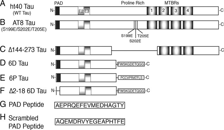
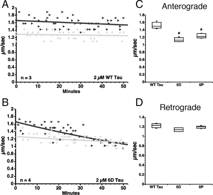
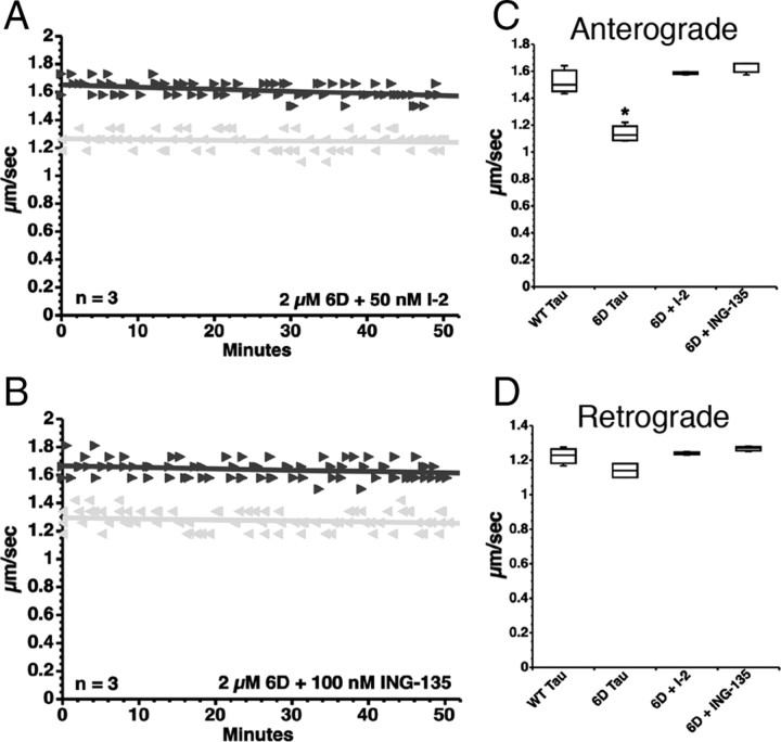
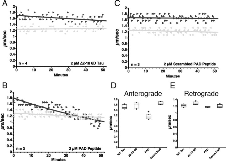
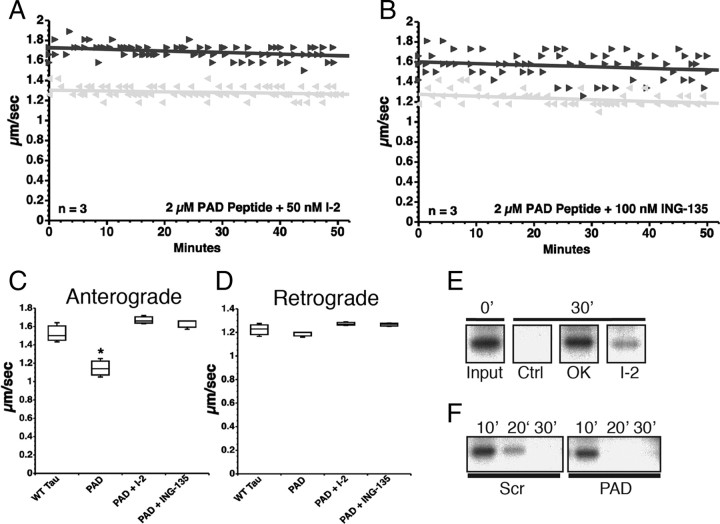
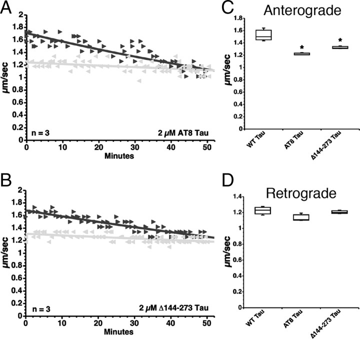
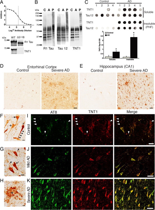
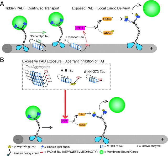
Aggregated filamentous forms of hyperphosphorylated tau (a microtubule-associated protein) represent pathological hallmarks of Alzheimer's disease (AD) and other tauopathies. While axonal transport dysfunction is thought to represent a primary pathogenic factor in AD and other neurodegenerative diseases, the direct molecular link between pathogenic forms of tau and deficits in axonal transport remain unclear. Recently, we demonstrated that filamentous, but not soluble, forms of wild-type tau inhibit anterograde, kinesin-based fast axonal transport (FAT) by activating axonal protein phosphatase 1 (PP1) and glycogen synthase kinase 3 (GSK3), independent of microtubule binding. Here, we demonstrate that amino acids 2-18 of tau, comprising a phosphatase-activating domain (PAD), are necessary and sufficient for activation of this pathway in axoplasms isolated from squid giant axons. Various pathogenic forms of tau displaying increased exposure of PAD inhibited anterograde FAT in squid axoplasm. Importantly, immunohistochemical studies using a novel PAD-specific monoclonal antibody in human postmortem tissue indicated that increased PAD exposure represents an early pathogenic event in AD that closely associates in time with AT8 immunoreactivity, an early marker of pathological tau. We propose a model of pathogenesis in which disease-associated changes in tau conformation lead to increased exposure of PAD, activation of PP1-GSK3, and inhibition of FAT. Results from these studies reveal a novel role for tau in modulating axonal phosphotransferases and provide a molecular basis for a toxic gain-of-function associated with pathogenic forms of tau.


Figures

Figure 1.
Figure 1.
Schematic diagram of the tau constructs used. A, Full-length wild-type ht40 (WT tau) is the largest isoform of tau found in the adult CNS. WT tau contains the PAD motif corresponding to amino acids 2–18 (black box), two alternatively spliced N-terminal exons (E2 and E3), and MTBRs 1–4. B, The AT8 tau protein was pseudophosphorylated at S199, S202, and T205 by mutagenesis of S → E and T → E to mimic the phosphoepitope recognized by the AT8 antibody (Biernat et al., 1992; Goedert et al., 1995) in hyperphosphorylated tau. C, The Δ144–273 tau mutant protein corresponds to a deletion of amino acids 144–273 (the proline-rich region and MTBR1) that was found in a patient with an autosomal-dominant familial form of FTD (Rovelet-Lecrux et al., 2009). D, The 6D tau protein is a noncanonical N-terminal isoform containing amino acids 1–143 with an additional 11 unique C-terminal amino acids (Luo et al., 2004). E, The 6P protein is the same as 6D, except the last 11 C-terminal amino acids are different (Luo et al., 2004). F, The Δ2–18 6D protein has amino acids 2–18 deleted from the N terminus. G, Synthetic peptide composed of the PAD sequence (amino acids 2–18 from WT tau). H, Synthetic peptide composed of the same amino acids as the PAD peptide, but rearranged in a random order.
Figure 2.
Figure 2.
N-terminal tau isoforms 6D and 6P tau selectively inhibit anterograde FAT. A–D, Vesicle motility assays in isolated squid axoplasm. Individual velocity (micrometers per second) rate measurements (arrowheads) are plotted as a function of time (in minutes). Black arrowheads and lines represent anterograde FAT rates (conventional kinesin-dependent), and gray arrows and lines represent retrograde FAT rates (cytoplasmic dynein-dependent). A, Perfusion with soluble wild-type tau monomers (WT tau) did not affect FAT rates in the anterograde (black triangle) or retrograde (gray triangle) direction. B, In contrast, soluble 6D tau monomers, which cannot bind microtubules, selectively inhibited the rate of anterograde FAT, while retrograde FAT was not significantly affected. C, Quantitative analysis of FAT demonstrates that 6D and 6P (n = 5) tau caused a significant reduction in anterograde FAT when compared with WT tau monomers (*p < 0.01, unpaired t test). D, Retrograde FAT was unaffected by 6D and 6P tau.
Figure 3.
Figure 3.
6D tau inhibits anterograde FAT via activation of a PP1–GSK3 cascade. A, Coperfusion of 6D tau and I-2, a specific PP1 inhibitor, prevented inhibition of anterograde (black triangle) FAT elicited by 6D (compare with Fig. 2B). B, Coperfusion of 6D tau and ING-135, a specific GSK3 inhibitor, similarly prevented inhibition of anterograde FAT. C, Quantitative analysis of FAT shows that inhibition of PP1 (via I-2) or inhibition of GSK3 (via ING-135) completely prevents the anterograde FAT inhibition elicited by 6D tau (one-way ANOVA, *p < 0.01 versus WT Tau, 6D+I-2, 6D+ING-135). D, Retrograde FAT (gray triangle, retrograde) was unaffected by 6D, regardless of whether inhibitors were present.
Figure 4.
Figure 4.
PAD is necessary and sufficient to trigger anterograde FAT inhibition. A, Perfusion of Δ2–18 6D into isolated axoplasm did not affect FAT in either direction (black triangle, anterograde; gray triangle, retrograde). B, In contrast, perfusion of the PAD peptide, which corresponds to amino acids 2–18 in WT tau, caused a reduction of anterograde, but not retrograde FAT rates. C, A scrambled version of the PAD peptide was used as a control, and when applied to squid axoplasms did not affect FAT. D, Quantitative analysis of FAT rates indicates that PAD peptide significantly inhibits anterograde FAT (one-way ANOVA, *p < 0.01), while deletion of the PAD sequence in 6D (Δ2–18 6D) and scrambling the PAD peptide (Scram PAD) completely prevented inhibition of anterograde FAT. E, Retrograde FAT rates were unaffected in all groups tested.
Figure 5.
Figure 5.
PAD peptide inhibits anterograde FAT by activation of the PP1–GSK3 cascade. A, Coperfusion of PAD peptide with I-2, a specific PP1 inhibitor, had no effect on anterograde (black triangle) or retrograde (gray triangle) FAT in squid axoplasm (compare with Fig. 4B). B, Coperfusion of PAD peptide with ING-135, a specific GSK3 inhibitor, similarly prevented inhibition of anterograde FAT by PAD. C, Quantitative analysis of FAT reveals that PAD peptide alone specifically and significantly inhibits anterograde FAT (one-way ANOVA, *p > 0.01), while addition of either PP1 or GSK3 inhibitors completely prevented the reduction in anterograde FAT rates induced by the PAD peptide. D, Retrograde FAT rates were unaffected in all groups tested. E, 32P-c-Jun (a phosphatase substrate) undergoes dephosphorylation by endogenous axoplasmic phosphatases after 30 min. Axoplasms were perfused with 32P-c-Jun alone (Ctrl) or with 32P-c-Jun and either okadaic acid (OK, 1 μm) or I-2 (400 nm). Aliquots were taken immediately after perfusion (0′) and 30 min later (30′), and analyzed by autoradiography. F, 32P-c-Jun was dephosphorylated to a greater extent in PAD-perfused axoplasms than its scrambled PAD-perfused “sister” counterpart, suggesting increased activation of endogenous axoplasmic phosphatases by PAD. Sister axoplasms from the same squid were perfused with 32P-c-Jun and either scrambled PAD (Scr) or PAD peptides, and analyzed by autoradiography (n = 2 sets of sister axoplasms).
Figure 6.
Figure 6.
Tau monomers with disease-associated modifications inhibit anterograde FAT. A, Perfusion of squid axoplasm with soluble tau monomers containing mutations S199E, S202E, and T205E (phosphomimicking the AT8 epitope seen in hyperphosphorylated tau; AT8 tau) causes a reduction in anterograde FAT (black triangle), but not retrograde (gray triangle) FAT. B, Similarly, perfusion of squid axoplasm with soluble Δ144–273 tau monomer (mimicking a deletion mutation associated with a case of FTD) results in a reduction in anterograde FAT, but not retrograde FAT. C, Quantitative analysis of FAT rates demonstrates that AT8 tau monomers and Δ144–273 tau monomers significantly inhibit anterograde FAT, when compared with WT tau monomers (one-way ANOVA, *p < 0.01). D, Retrograde FAT rates remained unaffected for all pathogenic tau monomers tested.
Figure 7.
Figure 7.
PAD immunoreactivity in AD brains. A, An ELISA titer of the TNT1 antibody using recombinant WT tau indicates a LogEC50 of 5.25 or a 1:180,000 dilution of a 1 mg/ml stock (final concentration, 5.5 ng/ml). TNT1 does not react with tau lacking PAD (amino acids 2–18; Δ2–18) in immunoblots. B, TNT1 labels enriched soluble tau from the frontal cortex of both control (Con Sol) and AD brains (AD Sol) as well as purified paired helical filament tau (AD PHF) from AD frontal cortex in denaturing conditions on Western blots. Comparison of TNT1 blots with Tau12 and R1 tau blots clearly indicates that TNT1 recognizes tau proteins and not other proteins in human brains. C, Dot blots of TNT1 under non-denaturing conditions. TNT1 preferentially labels soluble and PHF tau isolated from AD brains as opposed to tau isolated from control brains, indicating that PAD-exposed tau conformations are more abundant in AD. Quantification of both soluble and insoluble tau fractions is depicted as TNT1:Tau12 optical density (OD) ratios in the bar graph; Tau12 was used to measure total tau. *p ≤ 0.05, unpaired t test. D, E, TNT1 immunohistochemistry in the entorhinal cortex (D) and hippocampus (CA1 region; E) of human Braak stages I–II (control) or V–VI (severe AD) cases. Spatial and temporal patterns of TNT1 staining followed those of Braak staging, starting in the entorhinal cortex (D) and then appearing in the hippocampus (E) before involving temporal cortical gyri in later stages (data not shown). Scale bars, 100 μm. F, TNT1 staining in Braak stage I–II cases. TNT1 diffusely labeled the cytoplasm of pretangle CA2 neurons in the very early stages of inclusion formation (arrow). A subset of diffusely stained neurons contained globular cytoplasmic inclusions (filled arrow). A few neurons contained strongly labeled seemingly less mature tau inclusions that extended into the basal (arrowhead) and apical (black triangle) dendrites. G, H, TNT1 in Braak stages V–VI; note that large amounts of the classical tau inclusions, neurofibrillary tangles (arrow; G), neuropil threads (black triangle; G) and neuritic plaques (*; H), were present in the regions analyzed (CA1 shown). Scale bars, 20 μm. I–K, Representative images of TNT1 (red) and AT8 (green) double-label immunofluorescence in the hippocampus (CA1/CA2 region). I, The TNT1 epitope appears to precede that of AT8 in controls cases (Braak stages I–II). J, Both TNT1 and AT8 continue to accumulate in mild AD (Braak stages III–IV). K, TNT1 and AT8 appear with nearly complete colocalization in severe AD (Braak stages V–VI). Note that when present, AT8 extensively colocalizes with TNT1 at all Braak stages. Scale bars, 50 μm.
Figure 8.
Figure 8.
Proposed role of PAD in anterograde fast axonal transport regulation and in axonal transport dysfunction in disease. A, Tau is normally localized to microtubules where, in addition to stabilizing microtubules, it may regulate local cargo delivery through PAD-mediated activation of the PP1–GSK3 cascade. When tau is folded into the paperclip conformation, membrane-bound cargoes are transported by conventional kinesin along microtubules because PAD is hidden. In contrast, cargo is delivered at appropriate locations where tau is in an extended conformation and PAD is exposed. The exposure of PAD activates PP1-GSK3, leading to phosphorylation of kinesin light chains and release of cargoes. B, In disease states, tau aggregations and/or modifications expose PADs in tau that no longer binds to microtubules, promoting aberrant activation of the PP1–GSK3 cascade, increased inhibition of anterograde FAT, and ultimately neuron dysfunction/degeneration. Abnormal presentation of the biologically active PAD motif represents a critical factor in determining whether disease-associated forms of tau inhibit anterograde FAT.

References

    1. Alonso AC, Grundke-Iqbal I, Iqbal K. Alzheimer's disease hyperphosphorylated tau sequesters normal tau into tangles of filaments and disassembles microtubules. Nat Med. 1996;2:783–787. - PubMed
    1. Andreadis A. Tau gene alternative splicing: expression patterns, regulation and modulation of function in normal brain and neurodegenerative diseases. Biochim Biophys Acta. 2005;1739:91–103. - PubMed
    1. Arriagada PV, Growdon JH, Hedley-Whyte ET, Hyman BT. Neurofibrillary tangles but not senile plaques parallel duration and severity of Alzheimer's disease. Neurology. 1992;42:631–639. - PubMed
    1. Barghorn S, Davies P, Mandelkow E. Tau paired helical filaments from Alzheimer's disease brain and assembled in vitro are based on beta-structure in the core domain. Biochemistry. 2004;43:1694–1703. - PubMed
    1. Berry RW, Sweet AP, Clark FA, Lagalwar S, Lapin BR, Wang T, Topgi S, Guillozet-Bongaarts AL, Cochran EJ, Bigio EH, Binder LI. Tau epitope display in progressive supranuclear palsy and corticobasal degeneration. J Neurocytol. 2004;33:287–295. - PubMed

Publication types

MeSH terms

LinkOut - more resources