. 2011 Jul 6;31(27):9933-44.
doi: 10.1523/JNEUROSCI.1062-11.2011.

Primary cilia regulate proliferation of amplifying progenitors in adult hippocampus: implications for learning and memory

Affiliations

Primary cilia regulate proliferation of amplifying progenitors in adult hippocampus: implications for learning and memory

Alejandro Amador-Arjona et al. J Neurosci. .

Abstract

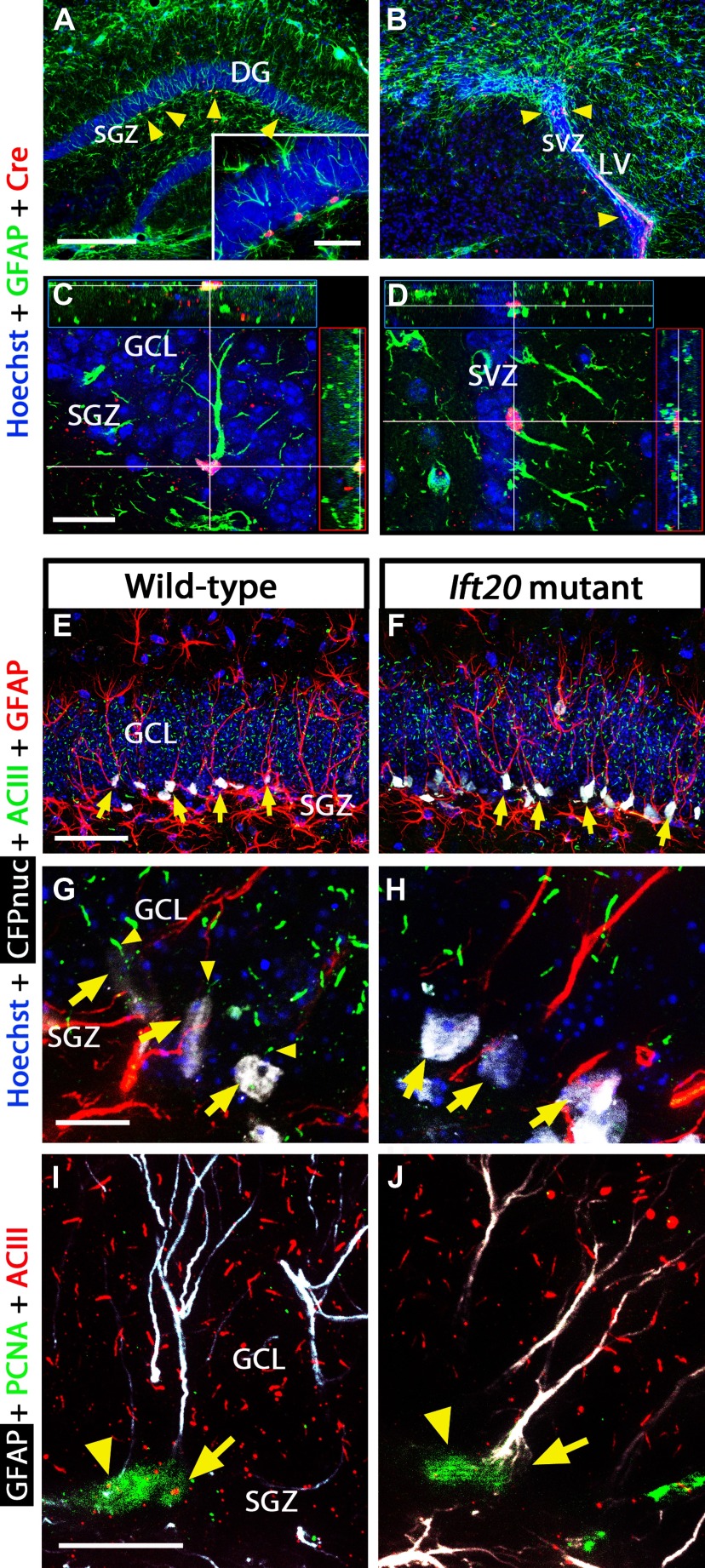
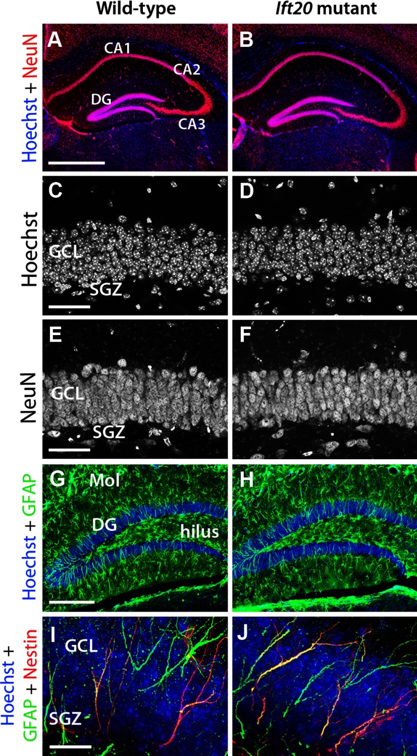
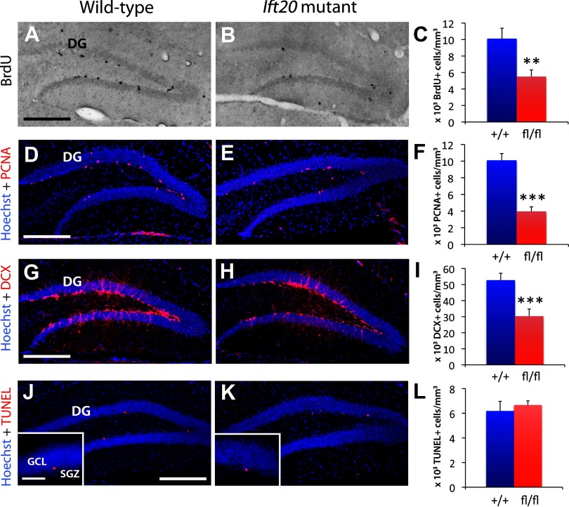
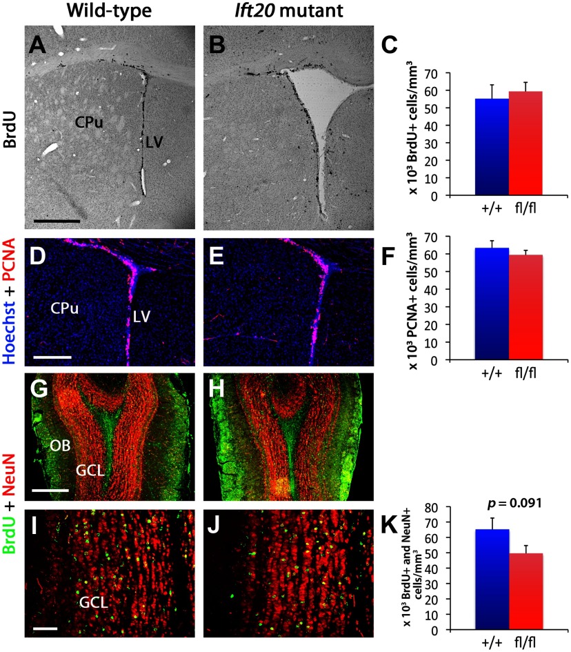
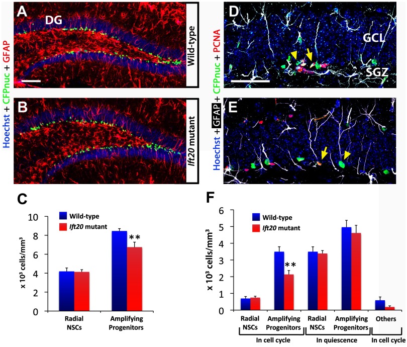
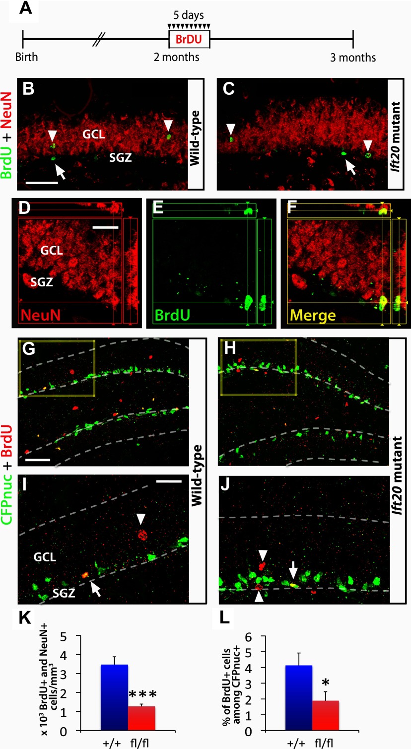
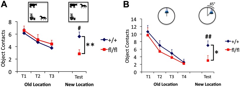
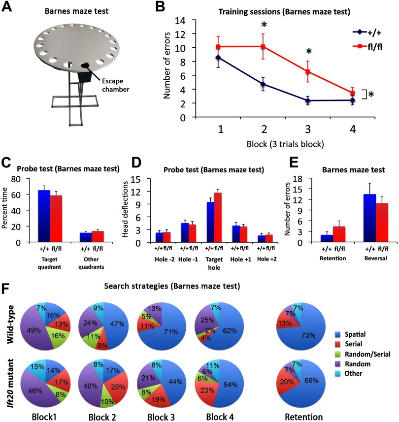
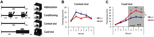
Integration of new neurons into the adult hippocampus has been linked to specific types of learning. Primary cilia were found to be required for the formation of adult neural stem cells (NSCs) in the hippocampal dentate gyrus during development. However, the requirement of cilia in maintenance of adult NSCs is unknown. We developed a genetic mouse model in which fetal/perinatal brain development is unaffected, but adult hippocampal neurogenesis is constantly reduced by conditional ablation of primary cilia in adult GFAP(+) neural stem/progenitor cells. We found that this approach specifically reduces the number of hippocampal amplifying progenitors (also called type 2a cells) without affecting the number of radial NSCs (or type 1 cells). Constant reduction of adult hippocampal neurogenesis produced a delay rather than a permanent deficiency in spatial learning without affecting the retention of long-term memories. Decreased neurogenesis also altered spatial novelty recognition and hippocampus-independent cue conditioning. Here, we propose that adult hippocampal newborn neurons increase the efficiency of generating the new representations of spatial memories and that reduction of adult hippocampal neurogenesis may be biased toward cue-based strategies. This novel mouse model provides evidences that cognitive deficits associated with ciliary defects (ciliopathies) might be, in part, mediated by the deficiency of primary cilia in adult hippocampal stem/progenitor cells.


Figures

Figure 1.
Figure 1.
Primary cilia are ablated in GFAP-expressing NSCs from Ift20fl/fl::mGFAP-Cre (Ift20 mutant) mice (Ift20+/+::mGFAP-Cre; wild type). Double immunostaining in 8-week-old Ift20 mutant mice shows Cre expression (red) in GFAP+ cells (green; arrowheads) in the SGZ (GCL, granule cell layer) of the hippocampal DG (A) and in the SVZ of the LVs (B). Confocal imaging showing GFAP+ cells expressing Cre in the SGZ (C) and in the SVZ (D) of 8-week-old Ift20 mutant mice. Confocal imaging with CFPnuc (white), ACIII (green; marker for primary cilia), and GFAP (red) in the DG showing primary cilia and GFAP-expressing quiescent NSCs (and hence are nestin-CFPnuc+; arrows) from 8-week-old Ift20+/+::mGFAP-Cre::nestin-CFPnuc (wild-type) (E) and Ift20fl/fl::mGFAP-Cre::nestin-CFPnuc (Ift20 mutant) (F) mice. Confocal imaging with CFPnuc (white), ACIII (green), and GFAP (red) in the SGZ of the DG showing primary cilia (arrowheads) in GFAP-expressing radial NSCs (arrows) from 8-week-old Ift20+/+::mGFAP-Cre::nestin-CFPnuc (wild type) (G), whereas equivalent cells do not show primary cilia in Ift20fl/fl::mGFAP-Cre::nestin-CFPnuc (Ift20 mutant) (H) mice. I, J, Confocal GFAP (white), PCNA (green), and ACIII (red) triple staining in DG of 8-week-old Ift20fl/fl::mGFAP-Cre (Ift20 mutant) and wild-type mice. Adult hippocampal stem/progenitor cells in cell cycle (PCNA+) show primary cilia staining (ACIII+) in wild-type but not in Ift20 mutants [arrows, radial NSCs (radial GFAP+); arrowheads, other cells in cell cycle]. Section thickness: A–J, 30 μm. Scale bars: A, B, 200 μm; C, D, I, J, 20 μm; E, F, 50 μm; G, H, 10 μm.
Figure 2.
Figure 2.
Neuromorphological characterization of the DG in 8-week-old Ift20 mutant (right panels) and wild-type (left panels) mice. A, B, NeuN (red) immunofluorescence shows preserved layering in hippocampal subregions (CA1, CA2, CA3, and DG) in Ift20 mutant and wild-type mice. C, D, Hoechst (white) nuclear staining in the GCL of the DG reveals no differences between Ift20 mutant and wild-type mice. E, F, NeuN (white) staining shows similar neuronal density in the GCL in both genotypes. G, H, Ift20 mutants show similar patterns of GFAP+ (green) astrocytes in the DG, hilus, and molecular layer (Mol) of the DG compared with wild-type mice. I, J, GFAP (green) and NESTIN (red) coexpression in cells from the SGZ of the DG indicates conserved morphology and distribution along the GCL. Section thickness: A–J, 30 μm. Scale bars: A, B, 500 μm; C–F, 50 μm; G, H, 200 μm; I, J, 20 μm.
Figure 3.
Figure 3.
Ift20 mutant mice exhibit reduced neural precursor cell proliferation in the SGZ of the DG. A, B, BrdU (black) staining after a 2 h BrdU pulse (100 mg kg−1 body weight) showed less proliferation in the SGZ of the DG in 8-week-old Ift20 mutants than in wild-type mice (quantified in C). D, E, Staining with the proliferative marker PCNA (red) confirmed that 8-week-old Ift20 mutants have approximately one-half the number of proliferating cells in the DG as wild-type mice. F, Quantification of PCNA+ cells. G, H, DCX-positive cell population (red) is reduced in the DG of 8-week-old Ift20 mutants. I, Quantification of DCX+ cells. J, K, Apoptosis (red) in the DG, as evaluated using the ApopTag Red In Situ Apoptosis Detection Kit (Millipore Bioscience Research Reagents), was not affected in 8-week-old Ift20 mutants relative to wild-type mice. L, Quantification of apoptotic cells. Section thickness: A, B, D, E, G, H, J, K, 30 μm. Scale bars: A, B, D, E, G, H, J, K, 200 μm; J, K, inset, 50 μm. **p < 0.01; ***p < 0.001. Data are expressed as mean values ± SEM.
Figure 4.
Figure 4.
Proliferation in the SVZ of the LVs is similar in Ift20 mutant (right panels) and wild-type mice (left panels). A, B, BrdU staining in 8-week-old mice after a 2 h BrdU pulse (100 mg kg−1 body weight) showed no evident genotypic differences in proliferation in the SVZ (CPu, caudate–putamen) (quantified in C). D, E, Staining with the proliferative marker PCNA (red) confirmed that proliferation in the SVZ is similar in Ift20 mutants and wild-type mice (quantified in F). G, H, A 5 d BrdU injection protocol (twice daily, 12 h apart, at 50 mg kg−1 body weight) was used in 8-week-old Ift20 mutant and wild-type mice. Double labeling of BrdU (green) and NeuN (red) in the olfactory bulb (OB) 4 weeks after BrdU injection in Ift20 mutant and wild-type mice shows similar distribution of adult newborn neurons in the GCL in the two groups. I, J, BrdU (green) and NeuN (red) double staining in the GCL of the OB 4 weeks after BrdU injection showed no genotypic differences in neurogenesis (quantified in K). Section thickness: A, B, D, E, G–J, 30 μm. Scale bars: A, B, 500 μm; D, E, G, H, 200 μm; I, J, 50 μm. Data are expressed as mean values ± SEM.
Figure 5.
Figure 5.
Number of adult hippocampal amplifying progenitors in cell cycle is reduced in Ift20fl/fl::mGFAP-Cre::nestin-CFPnuc (Ift20 mutant) mice. A, B, Confocal GFAP (red) and CFPnuc (green) double staining in DG of 8-week-old Ift20fl/fl::mGFAP-Cre::nestin-CFPnuc (Ift20 mutant) and Ift20+/+::mGFAP-Cre::nestin-CFPnuc (wild-type) mice allows the identification of radial NSCs that present a radial GFAP process and nuclear CFPnuc+ staining, and amplifying progenitors that present nuclear CFPnuc+ staining but without radial GFAP process. C, Quantification of radial NSCs (radial GFAP+) and amplifying progenitors (without GFAP+ radial process) CFPnuc+ cells revealed a reduction of amplifying progenitors. D, E, Confocal GFAP (white), CFPnuc (green), and PCNA (red) triple staining in DG of 8-week-old Ift20fl/fl::mGFAP-Cre::nestin-CFPnuc mice. D, Amplifying progenitor cells in cell cycle (arrow; PCNA+) and in quiescence (arrowhead; PCNA). E, Radial NSCs in cell cycle (arrow; PCNA+) and in quiescence (arrowhead; PCNA). F, Quantification of SGZ stem/progenitor cells (CFPnuc+) in 8-week-old Ift20fl/fl::mGFAP-Cre::nestin-CFPnuc (Ift20 mutant) and Ift20+/+::mGFAP-Cre::nestin-CFPnuc (wild-type) mice showed a decrease in number of amplifying progenitors in cell cycle (PCNA+). Radial NSCs and amplifying progenitor cells were distinguished by the presence or lack of a radial GFAP+ process, respectively. Section thickness: A, B, D, E, 30 μm. Scale bars: A, B, 100 μm; D, E, 25 μm. **p < 0.01. Data are expressed as mean values ± SEM.
Figure 6.
Figure 6.
Adult hippocampal neurogenesis is reduced in Ift20 mutant mice. A, A 5 d BrdU injection protocol (twice daily, 12 h apart, at 50 mg kg−1 body weight) was used in 8-week-old Ift20fl/fl::mGFAP-Cre (Ift20 mutant) and Ift20+/+::mGFAP-Cre (wild-type) mice, and the BrdU-labeled cells were examined 4 weeks after the last injection. B, C, BrdU (green) and NeuN (red) double staining 4 weeks after BrdU injection in Ift20 mutant and wild-type mice reveals BrdU-labeled adult newborn neurons (arrowheads; NeuN+) as well as other cell types in the SGZ (arrows; NeuN). D–F, Colocalization of a BrdU+ (green) cell that coexpress the neuronal marker NeuN (red). G, H, CFPnuc (green) and BrdU (red) double staining, 4 weeks after BrdU injection, in 8-week-old Ift20fl/fl::mGFAP-Cre::nestin-CFPnuc (Ift20 mutant) and Ift20+/+::mGFAP-Cre::nestin-CFPnuc (wild-type) mice showing colocalization of BrdU+/CFPnuc+ cells along the SGZ. Higher magnification (squares) of G and H are displayed in I and J, respectively (arrows, BrdU+ stem/progenitor cells; arrowheads, other BrdU+ cells). K, Quantification of BrdU+/NeuN+ cells showed reduced neurogenesis in the GCL of the DG in Ift20 mutants compared with wild-type mice. L, Quantification of the percentage of BrdU+ among CFPnuc+ cells indicated reduced numbers of stem/progenitor cells that retained BrdU in Ift20 mutants compared with wild-type mice. Section thickness: B–J, 30 μm. Scale bars: B, C, 100 μm; D–F, I, J, 25 μm; G, H, 50 μm. *p < 0.05; ***p < 0.001. Data are expressed as mean values ± SEM.
Figure 7.
Figure 7.
Ift20 mutant mice exhibit altered spatial novelty recognition. A, In a spatial pattern recognition test (mice group 1), habituation to three plastic objects (farmer boy, horse, and cow) across three familiarization trials (T1, T2, and T3) was similar in Ift20 mutant and wild-type mice, as they made fewer contacts with the objects over time. When one of the objects was moved to a novel location (test), a renewed interest (increased contacts) in the object was observed in wild-type but not in Ift20 mutant mice. B, In a location novelty recognition test (mice group 2), Ift20 mutants habituated to an object (Erlenmeyer flask filled two-thirds with water) across four familiarization trials (T1, T2, T3, and T4) similar as did wild type. Once the familiar object was shifted in an arc of 45° to a new location, a renewed interest (increased contacts) in the object was observed in wild type but not in Ift20 mutants. Group 1, Thirty-one mice (16 Ift20fl/fl::mGFAP-Cre and 15 wild-type); group 2, 21 mice (11 Ift20fl/fl::mGFAP-Cre and 10 wild-type). Differences between genotype were as follows: *p < 0.05; **p < 0.01. Differences between trial (old vs new location) were as follows: #p < 0.05; ##p < 0.01. Data are expressed as mean values ± SEM.
Figure 8.
Figure 8.
Ift20 mutant mice exhibit delayed spatial learning but preserved long-term memory. A, Schematic representation of Barnes maze test. B, In the Barnes maze, Ift20 mutants showed delayed spatial learning in finding the escape chamber (averaged into 4 blocks of 3 trials) but reached similar levels on task performance in the last block. C, D, Ift20 mutants and wild-type mice spent similar amount of time in the target quadrant (C) and made similar numbers of head deflections at the target hole during the probe test (no escape chamber) of the Barnes maze (D), showing that both groups had learned the original position of the chamber. E, Ift20 mutants showed normal retention and reversal of the task 2 weeks later. F, Ift20 mutants exhibited a delay in using a spatial strategy as evidenced by an increase in use of this strategy at the third block, whereas wild-type mice begin using a spatial strategy at the second block. Two weeks after the last trial (retention test), no differences in the use of spatial strategy were observed between the two genotypes. Shown is the percentage of each search strategies used by Ift20 mutants and wild-type mice across blocks (averaged into 4 blocks of 3 trials) and retention test on the Barnes maze (values represent group means). Search strategies are defined as follows: spatial, reaching the escape tunnel with scores of ≤3 for both error and distance (number of holes between the first hole visited and the escape tunnel); nonspatial, serial, random, random/serial, and other strategies are described in Materials and Methods. N = 31 mice (16 Ift20fl/fl::mGFAP-Cre and 15 wild-type). *p < 0.05. Data are expressed as mean values ± SEM.
Figure 9.
Figure 9.
Ift20 mutant mice exhibit enhanced fear responses to cued but not contextual fear conditioning. A, A 4 d fear conditioning protocol was used to evaluate contextual and cued fear conditioning. B, The freezing response to the same context than conditioning does not differ between the two genotypes (context test, mice group 1 and 2). C, Ift20 mutants show a higher freezing response during the exposition to the tone plus light combination (cue) in a different context (cued test, mice groups 1 and 2). Group 1, Thirty-one mice (16 Ift20fl/fl::mGFAP-Cre and 15 wild type); group 2, 21 mice (11 Ift20fl/fl::mGFAP-Cre and 10 wild type). *p < 0.05; **p < 0.01. Data are expressed as mean values ± SEM.

References

    1. Ahn S, Joyner AL. In vivo analysis of quiescent adult neural stem cells responding to Sonic hedgehog. Nature. 2005;437:894–897. - PubMed
    1. Aimone JB, Wiles J, Gage FH. Computational influence of adult neurogenesis on memory encoding. Neuron. 2009;61:187–202. - PMC - PubMed
    1. Aimone JB, Deng W, Gage FH. Adult neurogenesis: integrating theories and separating functions. Trends Cogn Sci. 2010;14:325–337. - PMC - PubMed
    1. Bach ME, Hawkins RD, Osman M, Kandel ER, Mayford M. Impairment of spatial but not contextual memory in CaMKII mutant mice with a selective loss of hippocampal LTP in the range of the theta frequency. Cell. 1995;81:905–915. - PubMed
    1. Bachevalier J, Nemanic S. Memory for spatial location and object-place associations are differently processed by the hippocampal formation, parahippocampal areas TH/TF and perirhinal cortex. Hippocampus. 2008;18:64–80. - PubMed

Publication types

MeSH terms