. 2011 Jul 6;31(27):9945-57.
doi: 10.1523/JNEUROSCI.1802-11.2011.

Bone marrow transplantation augments the effect of brain- and spinal cord-directed adeno-associated virus 2/5 gene therapy by altering inflammation in the murine model of globoid-cell leukodystrophy

Affiliations

Bone marrow transplantation augments the effect of brain- and spinal cord-directed adeno-associated virus 2/5 gene therapy by altering inflammation in the murine model of globoid-cell leukodystrophy

Adarsh S Reddy et al. J Neurosci. .

Abstract

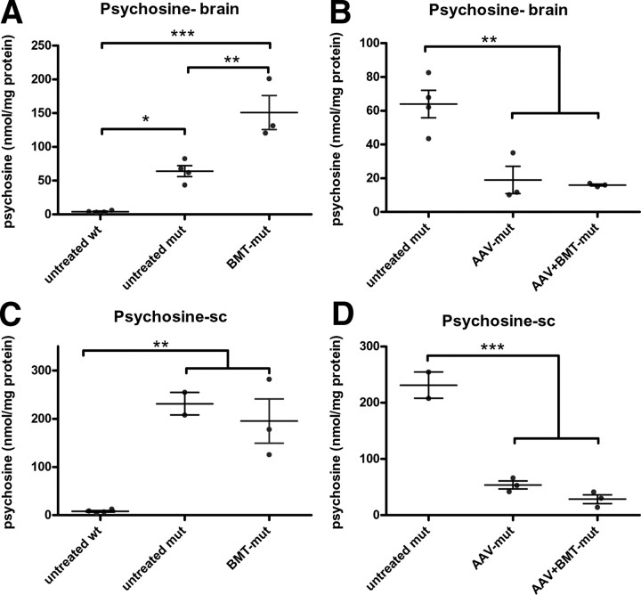
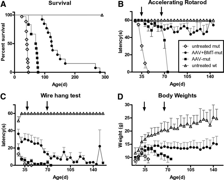
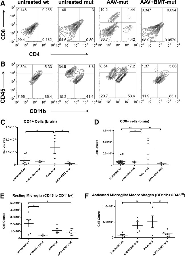
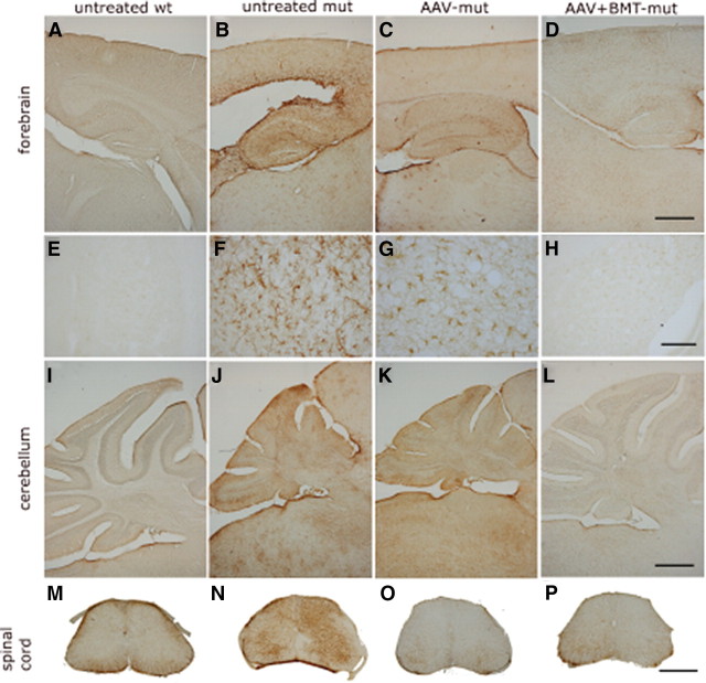
Globoid-cell leukodystrophy (GLD) is an inherited demyelinating disease caused by the deficiency of the lysosomal enzyme galactosylceramidase (GALC). A previous study in the murine model of GLD (twitcher) demonstrated a dramatic synergy between CNS-directed adeno-associated virus 2/5 (AAV2/5) gene therapy and myeloreductive bone marrow transplantation (BMT). However, the mechanism by which these two disparate therapeutic approaches synergize is not clear. In addition, the therapeutic efficacy may have been limited since the CNS-directed gene therapy was restricted to the forebrain and thalamus. In the current study, intrathecal and intracerebellar injections were added to the therapeutic regimen and the mechanism of synergy between BMT and gene therapy was determined. Although AAV2/5 alone provided supraphysiological levels of GALC activity and reduced psychosine levels in both the brain and spinal cord, it significantly increased CNS inflammation. Bone marrow transplantation alone provided essentially no GALC activity to the CNS and did not reduce psychosine levels. When AAV2/5 is combined with BMT, there are sustained improvements in motor function and the median life span is increased to 123 d (range, 92-282 d) compared with 41 d in the untreated twitcher mice. Interestingly, addition of BMT virtually eliminates both the disease and AAV2/5-associated inflammatory response. These data suggest that the efficacy of AAV2/5-mediated gene therapy is limited by the associated inflammatory response and BMT synergizes with AAV2/5 by modulating inflammation.


Figures

Figure 1.
Figure 1.
GALC activity and distribution. GALC activity was significantly higher in the untreated wt mice compared with the untreated mut and BMT-mut groups (A). There was no significant difference between the untreated mutant and BMT-mut groups (A). A separate analysis showed that the GALC activity in the brains of AAV-mut and AAV+BMT-mut groups was approximately fivefold greater and significantly different from that of the untreated wt group (B). The horizontal bars represent means, and error bars represent SEM. *p < 0.05; **p < 0.01; ***p < 0.001. GALC activity can be seen histochemically in the forebrain (C), cerebellum (G), and spinal cord (K) of the untreated wt group. In contrast, no GALC staining is observed in the forebrain (D), cerebellum (H), or spinal cord (L) of the untreated mut group. Intense GALC staining is observed in the ependyma of lateral and fourth ventricles of the AAV-mut and AAV+BMT-mut groups (asterisks; E, F, I, J). Intense staining was also observed in the meninges of the spinal cord and along the spinal nerve roots (arrows; M, N) of AAV-mut and AAV+BMT-mut mice. C–J were imaged at the same magnification. Scale bars: F, J, ∼600 μm. K–N were imaged at the same magnification. Scale bar: N, ∼600 μm.
Figure 2.
Figure 2.
Engraftment and GFP+ cells in the brain. The number of GFP+ donor cells present in the bone marrow and brains were determined using flow cytometry. The levels of bone marrow engraftment at day 36 were between 3 and 29%. There is no significant difference in donor engraftment in various groups receiving BMT (A). The number of cells present in FL1 channel (“GFP channel”) in the brain was similar in all the groups tested. There is no significant difference in the GFP+ cells in the brains between the treated and untreated groups (B). Error bars indicate SEM.
Figure 3.
Figure 3.
Psychosine levels. Psychosine levels in the brain (Psychosine-brain; A, B) and the spinal cord (Psychosine-sc; C, D) were measured using mass spectrometry. In the brain, the psychosine levels were highly elevated in the untreated mut compared with untreated wt group, while levels were significantly increased in the BMT-mut group relative to both the untreated wt and untreated mut groups (A). In spinal cord, both the untreated mut and BMT-mut groups had significantly increased psychosine levels compared with the untreated wt mice (C). The analyses conducted on treatments that decreased psychosine levels showed that both the AAV-mut and AAV+BMT-mut groups had significantly reduced levels of psychosine compared with the untreated mut mice in both brain (B) and spinal cord (D). There was no significant difference in psychosine levels between the AAV-mut and AAV+BMT-mut groups in either the brains or spinal cord. The horizontal bars represent the means, and the error bars represent SEM. *p < 0.05; **p < 0.01; ***p < 0.001.
Figure 4.
Figure 4.
LFB-PAS. Luxol fast blue/periodic acid–Schiff staining of the anterior commissure of the corpus callosum of representative animals is shown (A–D). The untreated wt animal (A) has essentially no PAS-positive macrophages within the white matter, the untreated mut (B) mouse shows prominent PAS-positive macrophages within the white matter (arrowheads), and both the AAV-mut (C) and AAV+BMT-mut (D) mice show a reduction in the number of PAS-positive macrophages in this region of the brain. Sections from the lateral white matter of the spinal cord from an untreated wt mouse (E) show no PAS-positive macrophages within the white matter, whereas sections from the same area of an untreated 36-d-old twitcher mouse (F) show numerous PAS-positive macrophages (arrowheads). There appears to be a slight reduction of PAS-positive cells in the spinal cords of both the AAV-mut (G) and AAV+BMT-mut groups (H).
Figure 5.
Figure 5.
Diffusion tensor imaging. Heat maps of axial diffusivity (λ||; A–D) and radial diffusivity (λ⊥; E–H) in the spinal cord obtained by DTI. In the DWM (I), there is a significant decrease in the axial diffusion in the untreated mut group compared with the untreated wt group. The AAV-mut and AAV+BMT-mut groups show an increase in axial diffusion compared with the untreated mut group. In the VLWM (J), the axial diffusivity of the untreated mut is significantly decreased compared with the untreated wt and the treated groups. Radial diffusivity in the DWM (K) is significantly increased in the untreated mut compared with the untreated wt. The treated groups are intermediate between the untreated wt and untreated mut groups. In the VLWM (L), there is a significant increase in the radial diffusivity in the untreated mut compared with untreated wt. There is no significant difference between the untreated wt and the AAV-mut and AAV+BMT-mut groups. The horizontal bars represent the means, and the error bars represent SEM. *p < 0.05; **p < 0.01; ***p < 0.001; ns, not significant.
Figure 6.
Figure 6.
Survival and behavior. Kaplan–Meier curves showing the survival of various treatment groups (A). The median life span of the AAV-mut group (71 d; range, 46–78 d) was significantly greater than (p < 0.001) that of the untreated mut group (41 d; range, 24–46 d). The median life span of the AAV+BMT-mut group (123 d; range, 92–282 d) was significantly greater than that of the AAV-mut group (p < 0.001). Behavior was evaluated using constant speed rotarod and wire hang test. Animals in the AAV-mut and AAV+BMT-mut group performed significantly better than the untreated mut group in the accelerating (B) rotarod motor function tests at 35 d and at 70 d of age (arrows). There was a statistically significant improvement in latency on wire hang test only in the AAV+BMT-mut group (C) at day 35 of age. Body weights (D) in the AAV+BMT-mut group were significantly higher compared with the untreated mut at 35 d of age. There was no significant difference between the AAV-mut and untreated mut groups at 35 d of age. At 70 d of age, the body weights were significantly higher in the AAV+BMT-mut group compared with the AAV-mut group. The body weights were maintained in the long-lived animals from the AAV+BMT-mut group. Error bars represent SEM.
Figure 7.
Figure 7.
Tremor. The averaged power spectrum (A) of the untreated mut group (blue line) is shifted toward higher frequencies with a broader bandwidth compared with the untreated wt group (solid black line). In the AAV-mut group (red line), the averaged power spectrum is similar to that of the untreated wild type. In the AAV+BMT-mut group (solid gray line), the power spectrum is shifted toward higher frequencies compared with the AAV-mut group and is similar to the untreated mut group. Compared with the untreated wt group, there is a significant increase in the frequency of peak power (B) and the power between 13 and 20 Hz (C) in the AAV+BMT-mut group, but not the AAV-mut group. The distance traveled by the AAV-mut and AAV+BMT-mut groups (D) is significantly increased compared with the untreated mut group. BMT alone altered the tremor phenotype in the wild-type mouse. The averaged power spectrum (A) of the BMT-WT group (solid green line) is shifted upward and rightward compared with the untreated wt group (solid black line). The frequency of peak power (B) and the power between 13 and 20 Hz (C) were significantly increased in the BMT-WT group compared with the untreated wild-type group. The distance traveled by the BMT-WT group was significantly decreased compared with the untreated wt group (D). The horizontal bars represent the mean, and the error bars represent SEM. ***p < 0.001; **p < 0.01; *p < 0.05.
Figure 8.
Figure 8.
Flow cytometry: brains. Representative bivariate plots show the relative numbers of CD4+ and CD8+ T-cells (A) and activated microglia (CD45hiCD11bhi) (B) in brains of untreated wt, untreated mut, AAV-mut, and AAV+BMT-mut groups. Quantitation of T-cells shows that there is a significant increase in CD4 and CD8 T-cells (C, D) in the AAV-mut group compared with the untreated wt. There is no increase in CD4 and CD8 T-cells in the untreated mut and AAV+BMT-mut groups compared with the untreated wt group (B–D). There appears to be an increase in CD4, CD8, and activated microglia (CD45hiCD11bhi) in the AAV-mut group compared with other groups. Quantitation of resting microglia (CD45loCD11bhi) (E) shows that there is a significant decrease in these cell numbers in the untreated mut and AAV+BMT-mut groups compared with the untreated wt group. Quantitation of activated microglia (F) shows that there is a significant increase in these cell numbers in the AAV-mut group compared with the untreated wt or AAV+BMT-mut group. There is no significant difference between the activated microglial numbers between untreated wt and AAV+BMT-mut groups. The horizontal bars represent means, and the error bars represent SEM. *p < 0.05, **p < 0.01.
Figure 9.
Figure 9.
Chemokines and cytokines. There is a significant increase in KC (A) in the brains of the untreated mut animals compared with the untreated wt animals. The chemokine MCP-1 (B) is increased in the untreated mut group and is undetectable in the untreated wt group. In both the AAV-mut and AAV+BMT-mut groups, KC and MCP-1 levels are reduced similar to the untreated wt group. The BMT-mut group does not show a decrease in the above chemokines compared with the untreated mut group. The chemokine MIP-1β (C) is undetectable in the untreated mut group and is present in the AAV-mut and AAV+BMT-mut groups at levels comparable with the untreated wt group. The levels of MIP-1β in the BMT-mut group are similar to that of the untreated mut group. The cytokine TNF-α (D) is significantly decreased in the untreated mut group compared with the untreated wt group. In the AAV-mut and AAV+BMT-mut groups, the levels of TNF-α are similar to that of the untreated wt group. The levels of TNF-α in the BMT-mut group are similar to that of the untreated mut group. The cytokine IL-12(p40) (E) shows a trend similar to KC, with a significant increase in the untreated mut group compared with the untreated wt group. Levels of IL-12(p40) in the AAV-mut and AAV+BMT-mut groups are similar to the untreated wt group. The horizontal bars represent mean, and error bars represent SEM. *p < 0.05; **p < 0.01; ***p < 0.001; ns, not significant.
Figure 10.
Figure 10.
GFAP immunohistochemistry. Representative images of GFAP staining of brain and spinal cords are shown. Untreated mut animals have higher GFAP immunoreactivity in all the regions of the CNS (B, F, J, N) compared with untreated wt animals (A, E, I, M). Animals in the AAV-mut group (C, G, K, O) appear to have similar or slightly decreased GFAP staining compared with the untreated mut group. The AAV+BMT-mut group (D, H, L, P) has less intense GFAP staining compared with the untreated mut and AAV-mut groups. High-magnification images (E–H) from the cortex show a characteristic activated astrocyte morphology. The spinal cords of the untreated wt group have minimal GFAP staining (M) compared with the untreated mut group (N). The spinal cords of the AAV-mut (O) and AAV+BMT-mut (P) groups stain with similar intensity as that of the untreated wt group. A–D and I–P were imaged at same magnification. Scale bars: D, L, P, ∼600 μm. E–H were imaged at same magnification. Scale bar: H, ∼25 μm.
Figure 11.
Figure 11.
CD68 immunohistochemistry. CD68 staining of the forebrain (A–D), cerebellum (E–H), and the spinal cord (I–L) at 36 d of age show increased staining in the untreated mut group (B, F, J). In the AAV-mut cerebellum (G), the CD68 staining appears similar or increased in intensity to that of the untreated mut (F). Interestingly, there appears to be decreased staining in the spinal cord (K) and the forebrain (C). In the AAV+BMT-mut group, the CD68 staining is decreased in most regions and indistinguishable from untreated wt mice in certain regions. A–H were imaged at the same magnification. Scale bars: D, H, ∼600 μm. I–L were imaged at the same magnification. Scale bar: L, ∼600 μm.

Similar articles

Cited by

References

    1. Arnett HA, Mason J, Marino M, Suzuki K, Matsushima GK, Ting JP. TNF alpha promotes proliferation of oligodendrocyte progenitors and remyelination. Nat Neurosci. 2001;4:1116–1122. - PubMed
    1. Biswas S, LeVine SM. Substrate-reduction therapy enhances the benefits of bone marrow transplantation in young mice with globoid cell leukodystrophy. Pediatr Res. 2002;51:40–47. - PubMed
    1. Biswas S, Biesiada H, Williams TD, LeVine SM. Substrate reduction intervention by l-cycloserine in twitcher mice (globoid cell leukodystrophy) on a B6;CAST/Ei background. Neurosci Lett. 2003;347:33–36. - PubMed
    1. Boisvert WA, Santiago R, Curtiss LK, Terkeltaub RA. A leukocyte homologue of the IL-8 receptor CXCR-2 mediates the accumulation of macrophages in atherosclerotic lesions of LDL receptor-deficient mice. J Clin Invest. 1998;101:353–363. - PMC - PubMed
    1. Bruscia EM, Ziegler EC, Price JE, Weiner S, Egan ME, Krause DS. Engraftment of donor-derived epithelial cells in multiple organs following bone marrow transplantation into newborn mice. Stem Cells. 2006;24:2299–2308. - PubMed

Publication types

MeSH terms