. 2011 Jul 6;31(27):10101-14.
doi: 10.1523/JNEUROSCI.0521-11.2011.

Molecular microdomains in a sensory terminal, the vestibular calyx ending

Affiliations

Molecular microdomains in a sensory terminal, the vestibular calyx ending

Anna Lysakowski et al. J Neurosci. .

Abstract

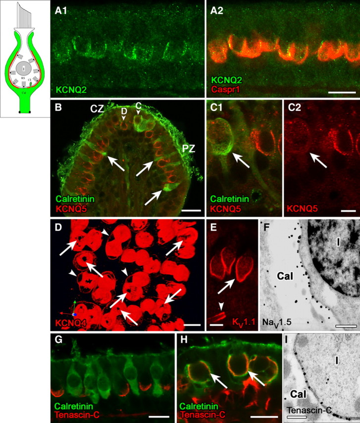
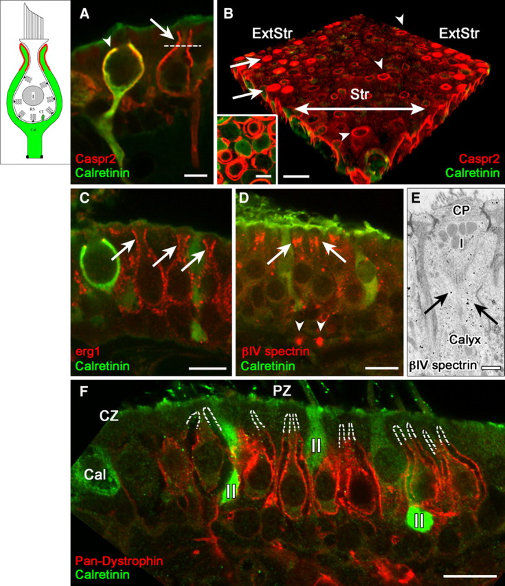
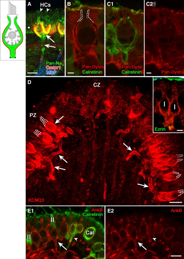
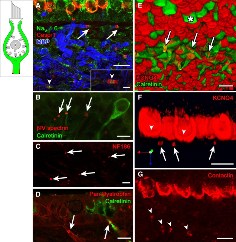
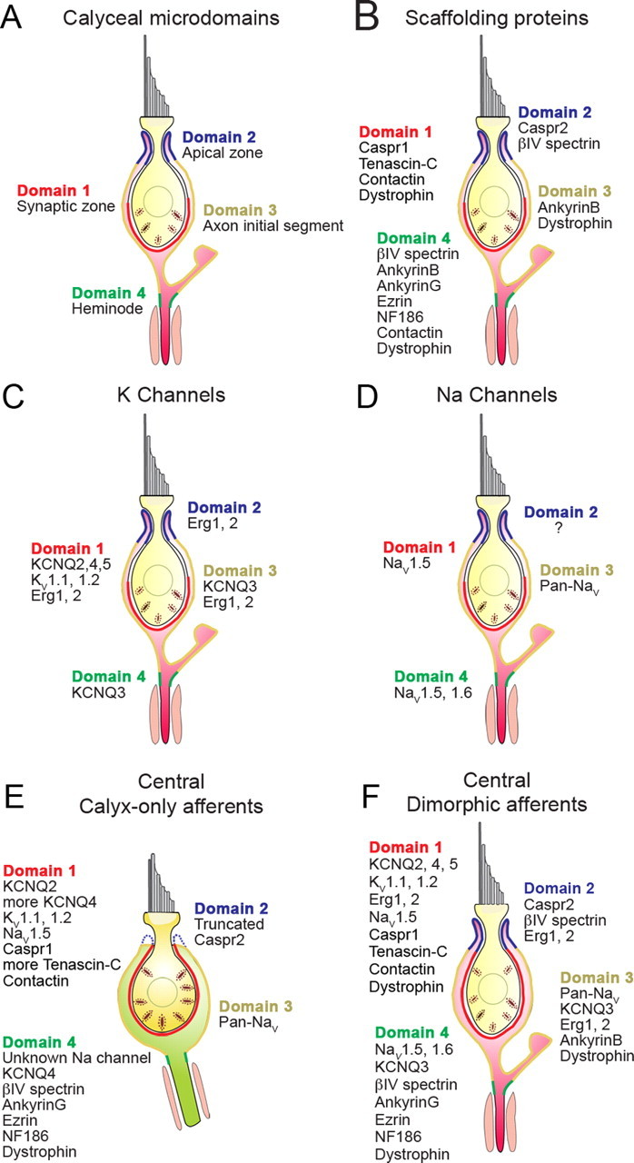
Many primary vestibular afferents form large cup-shaped postsynaptic terminals (calyces) that envelope the basolateral surfaces of type I hair cells. The calyceal terminals both respond to glutamate released from ribbon synapses in the type I cells and initiate spikes that propagate to the afferent's central terminals in the brainstem. The combination of synaptic and spike initiation functions in these unique sensory endings distinguishes them from the axonal nodes of central neurons and peripheral nerves, such as the sciatic nerve, which have provided most of our information about nodal specializations. We show that rat vestibular calyces express an unusual mix of voltage-gated Na and K channels and scaffolding, cell adhesion, and extracellular matrix proteins, which may hold the ion channels in place. Protein expression patterns form several microdomains within the calyx membrane: a synaptic domain facing the hair cell, the heminode abutting the first myelinated internode, and one or two intermediate domains. Differences in the expression and localization of proteins between afferent types and zones may contribute to known variations in afferent physiology.


Figures

Figure 1.
Figure 1.
Domain 1 of the calyx ending, facing the zone of presynaptic ribbons (RS) in the type I hair cell, is strongly immunoreactive for voltage-gated K and Na channels and associated proteins. Schematics in the upper left corner of Figures 1–4 delineate the successive domains in red. A, KCNQ2 immunostaining (A1, green) on the inner surface of the calyx ending, where it colocalized with Caspr1 (A2, red). B, C, KCNQ5 antibody (red) labeled the inner surface of calyces of both central-zone (CZ) and peripheral-zone (PZ) dimorphic (D) afferents (B, C1, C2, calretinin-negative), but not calyx-only (C) afferents (B, C1). Calretinin antibody (B, C1, green) selectively labels type II hair cells (arrows) and calyx-only (C) afferents, not dimorphic (D) afferents. Localization of the KCNQ5 label to the calyx membrane (rather than, e.g., the hair-cell membrane) was determined by immunogold EM (data not shown). D, Top view of a whole-mount utricular macula, stained with KCNQ4 antibody (red). The antibody stained the calyx membrane intensely on its inner face (domain 1) and lightly on its outer face (arrowheads, domain 3, see below). Interruptions in the KCNQ4 immunostaining at the bottom of each calyx appear as black holes (arrows). E, Antibodies to KV1.1 (red) and KV1.2 (similar pattern but not shown) also label domain 1 (arrow) in all calyces, in addition to labeling membrane of the nerve fiber near the first internode (the juxtaparaheminodal membrane, arrowhead). F, Immunoreactivity for NaV1.5 subunit was restricted to the calyx inner membrane, as shown here for a calyx (Cal) surrounding a type I hair cell (I); the calyx belonged to a dimorphic afferent in the peripheral zone of a crista. G, Antibody to tenascin-C (red) formed cup-like labeling at the base of type I hair cells, but not calretinin-positive type II hair cells (green) in the extrastriolar (peripheral) zone of the utricular macula. H, Tenascin-C forms much larger cups on these striolar calyces than on the extrastriolar calyces of G. Staining for tenascin-C (red) and calretinin (green) overlaps in confocal images, producing a yellow color in domain 1 (arrows) of calyx-only afferents in the striolar (central) zone. Tenascin-C antibody also labeled extracellular matrix in the tissue stroma below the sensory epithelium (see red stain below right-hand calyces). I, The higher resolution of immunogold EM revealed that tenascin-C is neither in the hair cell nor in the calyx, but rather in the synaptic cleft between the two (thus, the overlap in tenascin-C and calretinin in H reflects proximity of the antigens rather than colocalization, an underappreciated likelihood in most confocal studies). Shown are tenascin-C-immunolabeled gold particles in the synaptic cleft of a calyx in a crista peripheral zone. Such particles were not seen in the synaptic clefts of bouton synapses on type II hair cells (not shown). Scale bars: A, D, G, H, 10 μm; B, 20 μm; C, E, 5 μm; F, I, 500 nm.
Figure 2.
Figure 2.
Domain 2, the apical part of the calyx, was selectively labeled by antibodies to the cell adhesion molecule, Caspr2, and to erg KV subunits, and lacked several proteins found in domain 3. A, B, Caspr2 antibody (red) outlines domain 2: the apical part of the calyx (above the dashed line in A). These central calyces belong to a calyx-only (calretinin-positive) and a dimorphic (calretinin-negative) afferent. Domain 2 extends higher in dimorphic afferents (A, B, arrows) than in most calyx-only afferents (A, arrowhead), as shown in confocal transverse sections (A) and 3D reconstructions of whole-mount utricular maculae (B). In B, the striola (Str) had much less Caspr2 staining (red) than did the extrastriolar region (ExtStr). Arrowheads in B indicate Caspr2 label of three calyces of striolar dimorphic afferents, which extended to the apical surface of the epithelium. A transverse optical section of domain 2 (inset), taken above the level of the dotted line in A, shows concentric circles of Caspr2 label, demonstrating that it is present on both the inner and outer calyx surfaces. C, Domain 2 was labeled by antibodies to erg1 and erg2 (data not shown), but not by any of the other KV antibodies we used (KCNQ2, 3, 4, 5; KV1.1, 1.2). Erg1 antibody labeled domain 2 in calretinin-negative dimorphic afferents (arrows) but not in calyx-only afferents, as shown by the calretinin-positive (green) calyx on the left. Erg2 (data not shown) had a similar distribution. D, E, Immunoreactivity for βIV spectrin, which is usually found at nodes (arrowheads in D point to heminodes on the afferent fibers) and axonal initial segments, is also found in domain 2 (arrows), shown by both confocal immunohistochemistry (D) and EM immunogold localization. E, Note the gold particles (arrows) in the calyx surrounding the upper part of a type I (I) hair cell below the cuticular plate (CP). F, Domain 2 is also defined by the absence of several domain 3 proteins, including dystrophin (red); dashed white lines outline the unlabeled domain 2 in several calyces of peripheral-zone (PZ) dimorphic afferents. Dimorphic afferents are calretinin-negative, unlike central-zone (CZ) calyx-only afferents (green, Cal) and peripheral-zone (PZ) type II hair cells (II). Scale bars: A, B inset, 5 μm; B–D, F, 10 μm; E, 500 nm.
Figure 3.
Figure 3.
Domain 3, comprising the basolateral calyx and the stretch of axon leading to the heminode, selectively expressed NaV channels, KCNQ3, and certain scaffolding proteins. A, Pan-NaV antibody produced a complex pattern of label, including domains 1 and 3 (arrows) and the heminode (domain 4), as well as type I hair cells (arrowheads) and adjacent type II hair cells (asterisk). B, C, Pan-dystrophin (Pan-Dystr, red) labeled domain 3 of dimorphic (calretinin-negative), but not calyx-only (calretinin-positive, green) afferents (compare B with C1 and C2). D, Domain 3 in peripheral-zone dimorphic afferents was intensely immunoreactive for KCNQ3, as shown by this flattened projection of a stack of confocal images. KCNQ3 staining (arrows) extended throughout domain 3 from the basolateral outer surface of the calyx to the heminode. Note the lack of stain in domain 2, marked by dashed white lines. The inset shows a higher magnification single image of a dimorphic afferent with a “complex” calyx ending that envelops two adjacent type I hair cells. Antibody to ezrin (green) labeled the apical microvilli of the sensory epithelium and the heminode, where it combined with the red KCNQ3 immunolabel to form a yellow band. E, AnkyrinB antibody labeled domain 3 in dimorphic afferents (E1, E2, red, long arrows) but not calyx-only afferents (E1, E2, green, arrowheads). Scale bars: A, D, E, 10 μm; D, inset, 5 μm; B, C, 2 μm.
Figure 4.
Figure 4.
Domain 4, the heminodal membrane adjacent to the first internode, was marked by several ion channel and scaffolding proteins that mark nodes or initial segments in other tissues. A, Flattened projection of a confocal stack of images of the utricular epithelium and underlying stroma, where distal afferent fibers course before penetrating the basement membrane to approach the hair cells. Two heminodes (arrows) were labeled with the nodal markers Caspr1 (red) and NaV1.6 (green). They are located next to the first internodes, labeled by myelin basic protein (MBP, blue). Inset shows higher magnification of a full node (arrowhead, between two paranodes) from within the stroma, labeled with the same markers. Another full node is indicated in main panel (arrowhead). Heminodes (arrows) were also immunoreactive to antibodies against such other nodal markers as βIV spectrin (B), neurofascin186 (NF186, C), and pan-dystrophin (D). Note that the pan-dystrophin label in D encircles the cytoplasmic calretinin label of a calyx-only afferent, consistent with a membrane association. E, Three-dimensional reconstruction showing the underside of a whole-mount utricular macula. Striolar calyx-only afferents and extrastriolar type II hair cells (asterisk) are calretinin-positive (green). KCNQ4 antibody (red) labeled calyces and the heminodes (arrows) of striolar calyx-only afferents. The remaining structures are the inner calyx surfaces (domain 1), marked with dense KCNQ4 label (red) and viewed from below. F, A longitudinal section through striolar calyces from the same whole-mount utricular macula shown in Figure 1D, showing holes (arrowheads) in the bottom of the intense KCNQ4 label and several KCNQ4-positive heminodes (arrows). G, Contactin antibody (arrowheads) labeled paraheminodes in addition to domain 1. Comparison with F shows that heminodes are farther from the hair cell in these peripheral-zone afferents than in central-zone afferents. Scale bars: A–G, 10 μm; A, inset, 2.5 μm.
Figure 5.
Figure 5.
Ion channels and their associated proteins form calyceal microdomains in patterns that vary with afferent class and epithelial location. Most calyx endings belong to dimorphic afferents, which comprise 70–80% of vestibular afferents and innervate both central and peripheral zones of the epithelia. Domain 1 in peripheral/extrastriolar dimorphic afferents (A–D) neither extends as high nor contains as many synaptic ribbons (Lysakowski and Goldberg, 1997) as domain 1 in central/striolar afferents, whether calyx-only (E) or dimorphic (F). Domain 2 of peripheral dimorphic calyces is more extensive, corresponding to the longer, thinner necks of peripheral type I hair cells (Lysakowski and Goldberg, 1997). For the peripheral dimorphic calyces (A–D), we separately summarize calyceal microdomains (A), scaffolding proteins (B), KV channels (C), and NaV channels (D). E, Calyx-only afferents, which are restricted to central zones, appear to have a truncated domain 2 and do not express many of the proteins found in dimorphic calyces. F, In central dimorphic afferents, the synaptic zone (domain 1) extends farther upward than in peripheral dimorphic afferents. Central afferents of both types (E, F) express more KCNQ4 and tenascin-C than do peripheral dimorphic afferents.

References

    1. Abriel H, Kass RS. Regulation of the voltage-gated cardiac sodium channel Nav1.5 by interacting proteins. Trends Cardiovasc Med. 2005;15:35–40. - PubMed
    1. Arroyo EJ, Scherer SS. On the molecular architecture of myelinated fibers. Histochem Cell Biol. 2000;113:1–18. - PubMed
    1. Baird RA, Desmadryl G, Fernández C, Goldberg JM. The vestibular nerve of the chinchilla. II. Relation between afferent response properties and peripheral innervation patterns in the semicircular canals. J Neurophysiol. 1988;60:182–203. - PubMed
    1. Bal M, Zhang J, Zaika O, Hernandez CC, Shapiro MS. Homomeric and heteromeric assembly of KCNQ (Kv7) K+ channels assayed by total internal reflection fluorescence/fluorescence resonance energy transfer and patch clamp analysis. J Biol Chem. 2008;283:30668–30676. - PMC - PubMed
    1. Bao H, Wong WH, Goldberg JM, Eatock RA. Voltage-gated calcium channel currents in type I and type II hair cells isolated from the rat crista. J Neurophysiol. 2003;90:155–164. - PubMed

Publication types

MeSH terms

Substances

LinkOut - more resources