

Novel frontiers in ultra-structural and molecular MRI of the brain
- PMID: 21734576
- PMCID: PMC4217117
- DOI: 10.1097/WCO.0b013e328348972a
Novel frontiers in ultra-structural and molecular MRI of the brain
Abstract
Purpose of review: Recent developments in the MRI of the brain continue to expand its use in basic and clinical neuroscience. This review highlights some areas of recent progress.
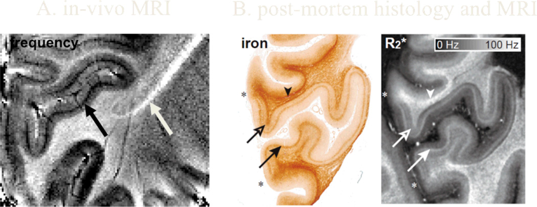
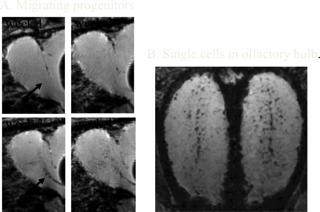
Recent findings: Higher magnetic field strengths and improved signal detectors have allowed improved visualization of the various properties of the brain, facilitating the anatomical definition of function-specific areas and their connections. For example, by sensitizing the MRI signal to the magnetic susceptibility of tissue, it is starting to become possible to reveal the laminar structure of the cortex and identify millimeter-scale fiber bundles. Using exogenous contrast agents, and innovative ways to manipulate contrast, it is becoming possible to highlight specific fiber tracts and cell populations. These techniques are bringing us closer to understanding the evolutionary blueprint of the brain, improving the detection and characterization of disease, and help to guide treatment.
Summary: Recent MRI techniques are leading to more detailed and more specific contrast in the study of the brain.
Figures
References
-
- Young IR, Hall AS, Pallis CA, Legg NJ, Bydder GM, Steiner RE. Nuclear magnetic resonance imaging of the brain in multiple sclerosis. Lancet. 1981;2(8255):1063–1066. - PubMed
-
- Bydder GM, Steiner RE, Young IR, Hall AS, Thomas DJ, Marshall J, Pallis CA, Legg NJ. Clinical nmr imaging of the brain. 140 cases. AJR Am J Roentgenol. 1982;139(2):215–236. - PubMed
-
- Ogawa S, Lee TM, Nayak AS, Glynn P. Oxygenation-sensitive contrast in magnetic resonance image of rodent brain at high magnetic fields. Magn Reson Med. 1990;14(1):68–78. - PubMed
-
- Le Bihan D. Looking into the functional architecture of the brain with diffusion mri. Nat Rev Neurosci. 2003;4(6):469–480. - PubMed
Publication types
MeSH terms
Grants and funding
LinkOut - more resources
Full Text Sources
Medical
