

Retinal amyloid peptides and complement factor H in transgenic models of Alzheimer's disease
- PMID: 21734608
- PMCID: PMC3719862
- DOI: 10.1097/WNR.0b013e3283497334
Retinal amyloid peptides and complement factor H in transgenic models of Alzheimer's disease
Abstract
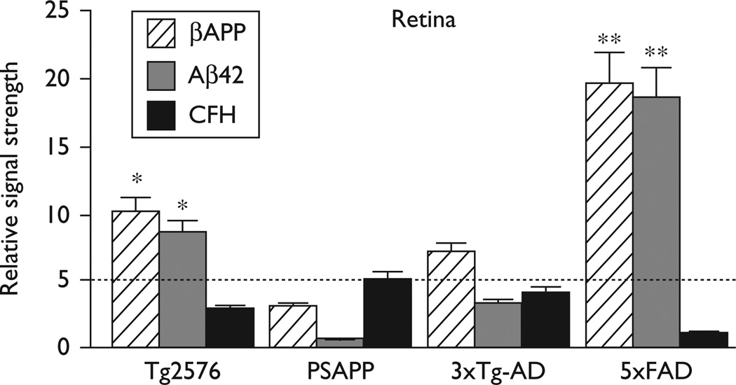
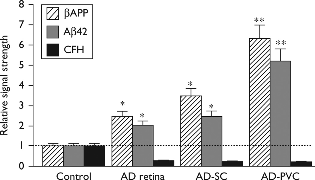
Murine transgenic models of Alzheimer's disease (Tg-AD) have been useful to analyze the contribution of β-amyloid precursor protein (βAPP), Aβ42 peptide deposition, and the proinflammatory mechanisms that characterize Alzheimer-type neuropathology. In this report, we have studied the levels of βAPP, Aβ40 and Aβ42 peptide, as well as the innate immune and inflammatory response-regulator complement factor H in the brain and retina in four different Tg-AD models including Tg2576, PSAPP, 3xTg-AD, and 5xFAD. Aged, symptomatic 5xFAD mice showed the highest retinal abundance of Aβ42 peptides and the highest deficits in complement factor H. This may be a useful model to study the mechanisms of amyloid-mediated inflammatory degeneration. The superior colliculus and retina obtained from late-stage Alzheimer's disease revealed upregulated amyloidogenic and inflammatory signaling along the anteroposterior axis of the retinal-primary visual cortex pathway.
Figures
References
-
- Philipson O, Lord A, Gumucio A, O’Callaghan P, Lannfelt L, Nilsson LN. Animal models of amyloid-beta-related pathologies in Alzheimer’s disease. FEBS J. 2010;277:1389–1409. - PubMed
-
- Veerhuis R. Histological and direct evidence for the role of complement in the neuroinflammation of Alzheimer’s disease. Curr Alzheimer Res. 2011;8:34–58. - PubMed
Publication types
MeSH terms
Substances
Grants and funding
LinkOut - more resources
Full Text Sources
Medical
Miscellaneous
