. 2011 Jul 6;475(7357):506-9.
doi: 10.1038/nature10307.

Legionella pneumophila SidD is a deAMPylase that modifies Rab1

Affiliations

Legionella pneumophila SidD is a deAMPylase that modifies Rab1

Yunhao Tan et al. Nature. .

Abstract

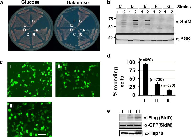
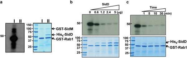
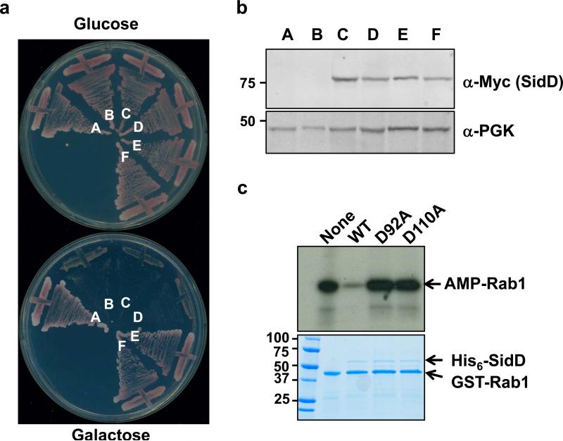
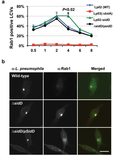
Legionella pneumophila actively modulates host vesicle trafficking pathways to facilitate its intracellular replication with effectors translocated by the Dot/Icm type IV secretion system (T4SS). The SidM/DrrA protein functions by locking the small GTPase Rab1 into an active form by its guanine nucleotide exchange factor (GEF) and AMPylation activity. Here we demonstrate that the L. pneumophila protein SidD preferably deAMPylates Rab1. We found that the deAMPylation activity of SidD could suppress the toxicity of SidM to yeast and is required to release Rab1 from bacterial phagosomes efficiently. A molecular mechanism for the temporal control of Rab1 activity in different phases of L. pneumophila infection is thus established. These observations indicate that AMPylation-mediated signal transduction is a reversible process regulated by specific enzymes.


Figures

Fig. 1
Fig. 1. Suppression of the cytotoxicity of SidM by SidD
a. Suppression of yeast toxicity of SidM. Yeast strains expressing SidM or SidM1-339 from a galactose-inducible promoter was transformed with various plasmids harboring sidD and the cells were streaked onto plates containing glucose or galactose. Plates were incubated at 30 °C for 3 days before acquiring the images. Yeast strains: A, vector/vector; B, vector/pSidD; C, pSidM/vector; D, pSidM/pSidD (original clone #1); E, pSidM/pSidD; F, pSidM1-339/vector; and G, pSidM1-339/pSidD. b. SidD did not affect the protein level of SidM or SidM1-339 in yeast cells. Subcultures of relevant yeast strains were grown in raffinose (1) or in galactose (2) medium. Crude lysates resolved by SDS-PAGE were probed with SidM-specific antibody. The 3-phosphoglycerate kinase (PGK) was used as a loading control (lower). c-e. Co-expression of SidD rescued the cell-rounding phenotypes caused by SidM1-339. 293T cells were transfected to express SidM1-339. A SidD plasmid was not included (I) or was used at 1:3 (II), or 1:5 (III) molar ratio, respectively. 24 hrs after transfection, samples were analyzed by acquiring images (c), by enumerating green cells exhibiting the rounding phenotype (d) or by immunoblotting to examine the protein levels of SidM1-339 and SidD (e). Experiments were repeated at three times and similar results were obtained. Error bars indicate s.d. Hsp70 was probed as a loading control. Bar, 50 μm.
Fig. 2
Fig. 2. SidD is a deAMPylase that targets SidM-modified Rab1
a. SidD prevented SidM-mediated AMPylation of Rab1. Shown were AMPylation reactions containing GSTSidM and GST-Rab1without (II) or with (I) His6-SidD. After SDS-PAGE, 32P-α-AMPRab1 was detected by autoradiography (left panel) and proteins were detected by Coomassie bright blue staining (right panel). b. Dose-dependent deAMPylation by SidD. His6-SidD was added to identical samples containing AMPylated GST-Rab1 to establish reactions in which the molar ratio of Rab1 and SidD is 8, 4, 2 and 1, respectively; reactions were terminated after 5 min of incubation. AMPylated GST-Rab1 and proteins in reactions were detected as described in a. c. Time course of SidD activity. AMPylated GST-Rab1 was mixed with His6-SidD at a molar ratio of 10:1, reactions were terminated at the indicated time intervals. AMPylated GST-Rab1 (upper panel) and proteins (lower) were similarly detected. Markers for protein size (kDa) are indicated.
Fig. 3
Fig. 3. The Asp residue at position 92 or 110 is important for SidD activity
a. Mutations of D92 and D110 abolished the ability of SidD to suppress the yeast cytotoxicity of SidM. Plasmids harboring sidD or its mutants were transformed into the SidM-expression yeast strain; cells were streaked onto plates containing glucose or galactose. Yeast strains: A, vector/vector; B, pSidM/vector; C, pSidM/pSidDD92A; D, pSidM/pSidDD110A; E, pSidM/pSidDD60A and F, pSidM/pSidD. b. Expression of the SidD mutants in yeast, samples prepared as described in Figure 1 and were probed for SidD (Myc-tagged) and for PGK. c. SidDD92A and SidDD110A have lost the deAMPylation activity. 1.5 micrograms purified proteins were added to reactions containing AMPylated Rab1. After 30 min incubation, reactions were terminated by SDS sample buffer. 32P-α-AMP-GST-Rab1 was detected by autoradiography and the proteins were detected by Coomassie bright blue staining (lower panel). Protein size (kDa) references are indicated on the left lane of the gel.
Fig. 4
Fig. 4. SidD is required for efficient removal of Rab1 from L. pneumophila phagosome
a. Mouse macrophages were infected with relevant L. pneumophila strains. At the indicated time points, fixed samples were probed for L. pneumophila and Rab1 with specific antibodies followed by Texas red and FITC-conjugated secondary antibodies, respectively. Processed samples were scored for co-localization of Rab1 with the bacterial phagosomes. Data shown are from two independent experiments performed in triplicate in which at least 100 phagosomes were scored per sample. b. Association of Rab1 with L. pneumophila phagosome 4 hrs after infection. Shown are images of wild type (Lp02, dot/icm+), the sidD deletion mutant (Lp02ΔsidD, dot/icm+) and the complementation strain (Lp02ΔsidD/Flag-SidD) residing in macrophages 4 hrs after infection. L. pneumophila and Rab1 are labeled as described in a, with bacteria marked in red and Rab1 marked in green. Bar, 10 μm. Note the difference in the intensity of Rab1 staining signals among the three bacterial strains. At least 150 vacuoles were scored each sample and error bars indicate s.d. Similar results were obtained in at least three independent experiments.

Comment in

References

    1. Ensminger AW, Isberg RR. Legionella pneumophila Dot/Icm translocated substrates: a sum of parts. Curr Opin Microbiol. 2009;12:67–73. - PMC - PubMed
    1. Machner MP, Isberg RR. Targeting of host Rab GTPase function by the intravacuolar pathogen Legionella pneumophila. Dev Cell. 2006;11:47–56. - PubMed
    1. Murata T, et al. The Legionella pneumophila effector protein DrrA is a Rab1 guanine nucleotide-exchange factor. Nat Cell Biol. 2006;8:971–977. - PubMed
    1. Muller MP, et al. The Legionella effector protein DrrA AMPylates the membrane traffic regulator Rab1b. Science. 2010;329:946–949. - PubMed
    1. Isberg RR, O'Connor TJ, Heidtman M. The Legionella pneumophila replication vacuole: making a cosy niche inside host cells. Nat Rev Microbiol. 2009;7:13–24. - PMC - PubMed

Publication types

MeSH terms

Substances