

Role of temporal processing stages by inferior temporal neurons in facial recognition
- PMID: 21734904
- PMCID: PMC3124819
- DOI: 10.3389/fpsyg.2011.00141
Role of temporal processing stages by inferior temporal neurons in facial recognition
Abstract
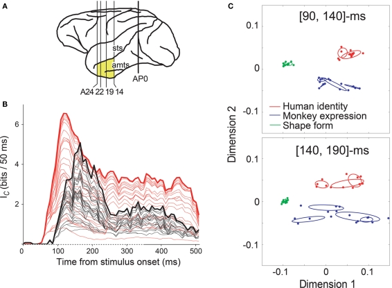
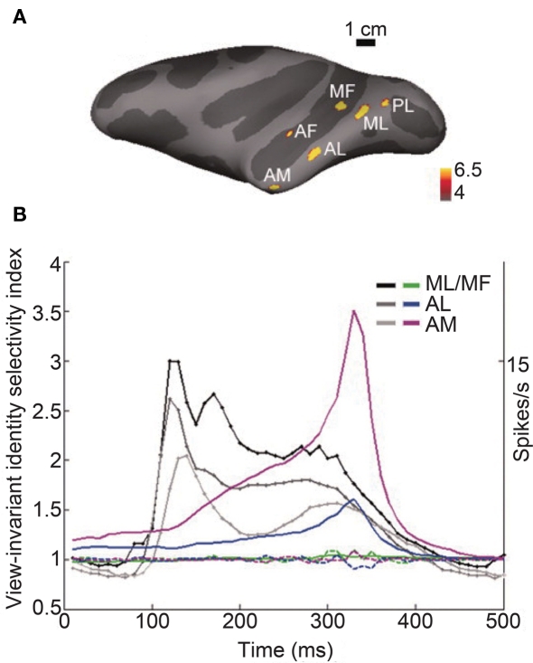
In this review, we focus on the role of temporal stages of encoded facial information in the visual system, which might enable the efficient determination of species, identity, and expression. Facial recognition is an important function of our brain and is known to be processed in the ventral visual pathway, where visual signals are processed through areas V1, V2, V4, and the inferior temporal (IT) cortex. In the IT cortex, neurons show selective responses to complex visual images such as faces, and at each stage along the pathway the stimulus selectivity of the neural responses becomes sharper, particularly in the later portion of the responses. In the IT cortex of the monkey, facial information is represented by different temporal stages of neural responses, as shown in our previous study: the initial transient response of face-responsive neurons represents information about global categories, i.e., human vs. monkey vs. simple shapes, whilst the later portion of these responses represents information about detailed facial categories, i.e., expression and/or identity. This suggests that the temporal stages of the neuronal firing pattern play an important role in the coding of visual stimuli, including faces. This type of coding may be a plausible mechanism underlying the temporal dynamics of recognition, including the process of detection/categorization followed by the identification of objects. Recent single-unit studies in monkeys have also provided evidence consistent with the important role of the temporal stages of encoded facial information. For example, view-invariant facial identity information is represented in the response at a later period within a region of face-selective neurons. Consistent with these findings, temporally modulated neural activity has also been observed in human studies. These results suggest a close correlation between the temporal processing stages of facial information by IT neurons and the temporal dynamics of face recognition.
Keywords: categorization; facial identity; inferior temporal cortex; temporal dynamics.
Figures
Similar articles
-
[Neural Mechanisms Underlying the Face Inversion Effect].Brain Nerve. 2015 Oct;67(10):1231-9. doi: 10.11477/mf.1416200288. Brain Nerve. 2015. PMID: 26450075 Review. Japanese.
-
Population dynamics of face-responsive neurons in the inferior temporal cortex.Cereb Cortex. 2005 Aug;15(8):1103-12. doi: 10.1093/cercor/bhh209. Epub 2004 Nov 24. Cereb Cortex. 2005. PMID: 15563724
-
Brain mechanisms for invariant visual recognition and learning.Behav Processes. 1994 Dec;33(1-2):113-38. doi: 10.1016/0376-6357(94)90062-0. Epub 2002 May 31. Behav Processes. 1994. PMID: 24925242
-
[Neural representations of facial identity and its associative meaning].Brain Nerve. 2012 Jul;64(7):841-52. Brain Nerve. 2012. PMID: 22764356 Review. Japanese.
-
Simple Learned Weighted Sums of Inferior Temporal Neuronal Firing Rates Accurately Predict Human Core Object Recognition Performance.J Neurosci. 2015 Sep 30;35(39):13402-18. doi: 10.1523/JNEUROSCI.5181-14.2015. J Neurosci. 2015. PMID: 26424887 Free PMC article.
Cited by
-
How the visual cortex handles stimulus noise: insights from amblyopia.PLoS One. 2013 Jun 20;8(6):e66583. doi: 10.1371/journal.pone.0066583. Print 2013. PLoS One. 2013. PMID: 23818947 Free PMC article. Clinical Trial.
-
Implications of holistic face processing in autism and schizophrenia.Front Psychol. 2013 Jul 5;4:414. doi: 10.3389/fpsyg.2013.00414. eCollection 2013. Front Psychol. 2013. PMID: 23847581 Free PMC article.
-
Enhanced intrinsic functional connectivity in the visual system of visual artist: Implications for creativity.Front Neurosci. 2023 Feb 22;17:1114771. doi: 10.3389/fnins.2023.1114771. eCollection 2023. Front Neurosci. 2023. PMID: 36908805 Free PMC article.
-
Structure supports function: Informing directed and dynamic functional connectivity with anatomical priors.Netw Neurosci. 2022 Jun 1;6(2):401-419. doi: 10.1162/netn_a_00218. eCollection 2022 Jun. Netw Neurosci. 2022. PMID: 35733424 Free PMC article.
-
Enhanced white matter fiber tract of the cortical visual system in visual artists: implications for creativity.Front Neurosci. 2023 Oct 25;17:1248266. doi: 10.3389/fnins.2023.1248266. eCollection 2023. Front Neurosci. 2023. PMID: 37946727 Free PMC article.
References
-
- Amaral D. G., Price J. L., Pitkanen A., Carmichael S. T. (1992). Anatomical Organization of the Primate Amygdaloid Complex. New York: Wiley-Liss, Inc
LinkOut - more resources
Full Text Sources